一句话亮点
这项研究发现,结直肠癌细胞在接受FOLFOX化疗时,细胞内的自噬不仅不是“帮倒忙”的耐药机制,反而像一个“免疫总指挥”——通过降解E3泛素连接酶FBXW2,稳定转录因子C/EBPβ,上调分泌蛋白TIMP-2,从而抑制MMP-2/9活性,把CD8⁺ T细胞“拉拢”到肿瘤里来“帮忙”,最终增强化疗疗效。这个“自噬→TIMP-2→T细胞”轴为解释化疗如何调动免疫系统提供了全新视角。
背景/痛点
FOLFOX(5-FU+奥沙利铂)是结直肠癌(CRC)的基石化疗方案,但耐药仍是临床核心难题。过去大家主要盯着化疗对肿瘤细胞的直接杀伤,但近年来发现,化疗的效果很大程度上依赖它能否重塑肿瘤免疫微环境(TIME)——尤其是能否把CD8⁺ T细胞招募进肿瘤、激活起来。
另一方面,自噬在癌症中的角色一直很“分裂”:在免疫缺陷小鼠(没T细胞)的模型中,抑制自噬反而增强化疗敏感性,说明自噬在肿瘤细胞内部有“保命”作用;但在免疫功能正常的小鼠中,抑制自噬却让化疗失效了。这意味着在真实的、有免疫系统的环境下,自噬可能干了一件更重要的事——调控抗肿瘤免疫。但具体怎么干的,机制不清。
这就引出了一个“反转”式的问题:化疗诱导的自噬,到底是“帮肿瘤抵抗”还是“帮免疫系统打肿瘤”?本文正是要解开这个结。
推理链分步拆解
第一步:自噬是“免疫依赖型”化疗敏感性的前提
他们先在体外用CRISPR把CT-26(小鼠CRC细胞)的ATG5或ATG7敲掉,确认自噬被彻底阻断。然后分别把野生型和自噬缺陷的肿瘤种到免疫缺陷裸鼠和免疫正常小鼠身上,用FOLFOX方案治疗。
结果很有意思:在裸鼠里,敲掉ATG5反而让化疗效果更好——这和传统“自噬促存活”的观点一致。但在免疫正常小鼠里,敲掉ATG5竟然让化疗失效了,肿瘤长得跟没治疗一样。用氯喹(CQ)药理学抑制自噬,也得到同样结果。
@方法论点评:“免疫缺陷 vs 免疫正常”的平行对比实验是区分“肿瘤细胞自主性”与“免疫依赖性”效应的黄金设计。如果只在裸鼠里做,就会得出“抑制自噬增敏化疗”的片面结论,甚至可能误导临床策略。这里的反转很关键,说明自噬对化疗的影响在免疫完整环境中完全被“免疫调节”功能盖过了。
那么问题来了:自噬到底改变了TIME里的什么?
第二步:自噬缺失,“拉拢”CD8⁺ T细胞的能力没了
他们用CyTOF(质谱流式)对肿瘤浸润免疫细胞做了全景式分析,结果很清晰:化疗后,野生型肿瘤里CD8⁺ T细胞和NK细胞明显增多,巨噬细胞减少;但ATG5敲除后,化疗引起的CD8⁺ T细胞扩增被特异性阻断,而NK细胞和巨噬细胞变化不大。
进一步分析CD8⁺ T细胞亚群,发现化疗诱导的Ly6C⁺ CD8⁺效应/记忆T细胞(高表达GZMB、穿孔素、TNFα,低表达PD-1/TIM-3)在自噬缺陷肿瘤中几乎消失。
他们还做了体外实验:取化疗后野生型肿瘤的间质液(TIF)去刺激CD8⁺ T细胞,能显著促进其增殖和杀伤功能;但自噬缺陷肿瘤的TIF就没有这个能力。
@方法论点评:用TIF替代肿瘤细胞直接共培养,能排除细胞接触依赖的信号,专门“钓”出分泌性因子。这是个典型的“条件培养基”逻辑,说明自噬是通过分泌某种可溶性因子来远程调控T细胞的,而不是通过细胞间直接接触。
那这个“神秘因子”是什么?
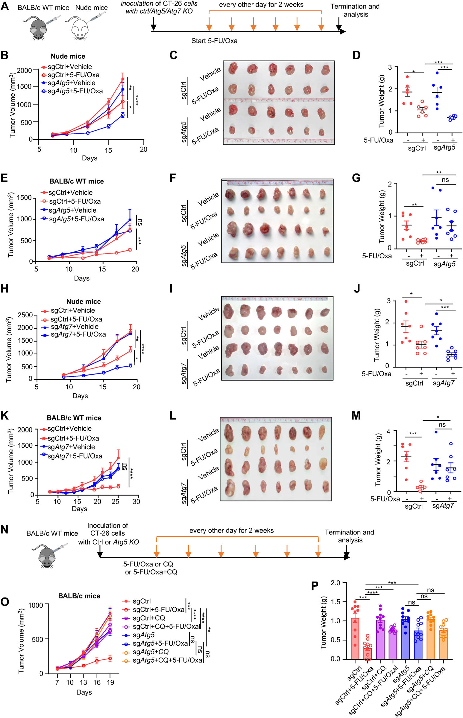
Fig. 1. Tumor cell-intrinsic autophagy is required for efficacy of chemotherapy in immunocompetent host. (A) Schematic representation of the treatment schedule in BALB/c nude and wild-type (WT) mice bearing subcutaneous CT-26 tumors with or without Atg5/Atg7 knockout (sgCtrl vs sg Atg5 /sg Atg7 ). All drugs (5-FU, 25 mg/kg; Oxa, 2.5 mg/kg) were administered intraperitoneally (i.p.) every other day for 2 weeks. (B–G) Growth curves of tumors, representative images, and tumor weights for CT-26 tumors with or without sg Atg5 in nude (B–D) or WT (E–G) mice (n = 6–7 per group). (H–M) Growth curves of tumors, representative images, and tumor weights for sgCtrl versus sg Atg7 tumors in nude (H–J) or WT (K–M) mice (n = 7 per group). (N) Treatment scheme for CT-26 tumors with or without sg Atg5 in WT mice for assessing the efficacy of 5-FU/Oxa, CQ alone, or in combination (n = 8-10 per group). All drugs were administered intraperitoneally (i.p.) every other day for 2 weeks; CQ was given at a dose of 60 mg/kg. (O) Tumor growth curves, and (P) tumor weights for the indicated treatment groups. Data are shown as mean ± SEM. Statistical analysis was performed using Two-way ANOVA with multiple comparisons. Significance is denoted as p < 0.05, p < 0.01, p < 0.001, p < 0.0001, ns, not significant.(图注取自PDF文本层,来源:Advanced Science, 2026)
第三步:锁定TIMP-2——自噬调控免疫的“关键信使”
他们用RNA-seq筛选化疗后受自噬调控的基因,发现“细胞因子相关通路”富集。于是直接用膜芯片筛查培养上清中的80种细胞因子,最终发现7个候选,其中TIMP-2引起了最大兴趣——因为TIMP-2是MMP-2/9的内源性抑制剂,而其表达水平在CRC中与预后相关,但在免疫调控中的角色几乎未知。
验证实验很干净:
敲除TIMP-2:在免疫正常小鼠中,TIMP-2 KO完全复制了自噬缺陷导致的化疗失效;但在裸鼠中,TIMP-2 KO对化疗没有影响——说明TIMP-2的作用是“免疫依赖”的。 在自噬缺陷细胞中回补TIMP-2:能显著恢复化疗敏感性,同时恢复CD8⁺ T细胞浸润。 用CD8⁺ T细胞清除抗体:回补TIMP-2带来的疗效恢复被完全逆转——证明TIMP-2是通过CD8⁺ T细胞来起作用的。
@方法论点评:“KO回补实验 + 清除实验”构成因果链闭环。先证明TIMP-2是自噬的下游,再证明其功能依赖于CD8⁺ T细胞,层层剥离,逻辑严密。
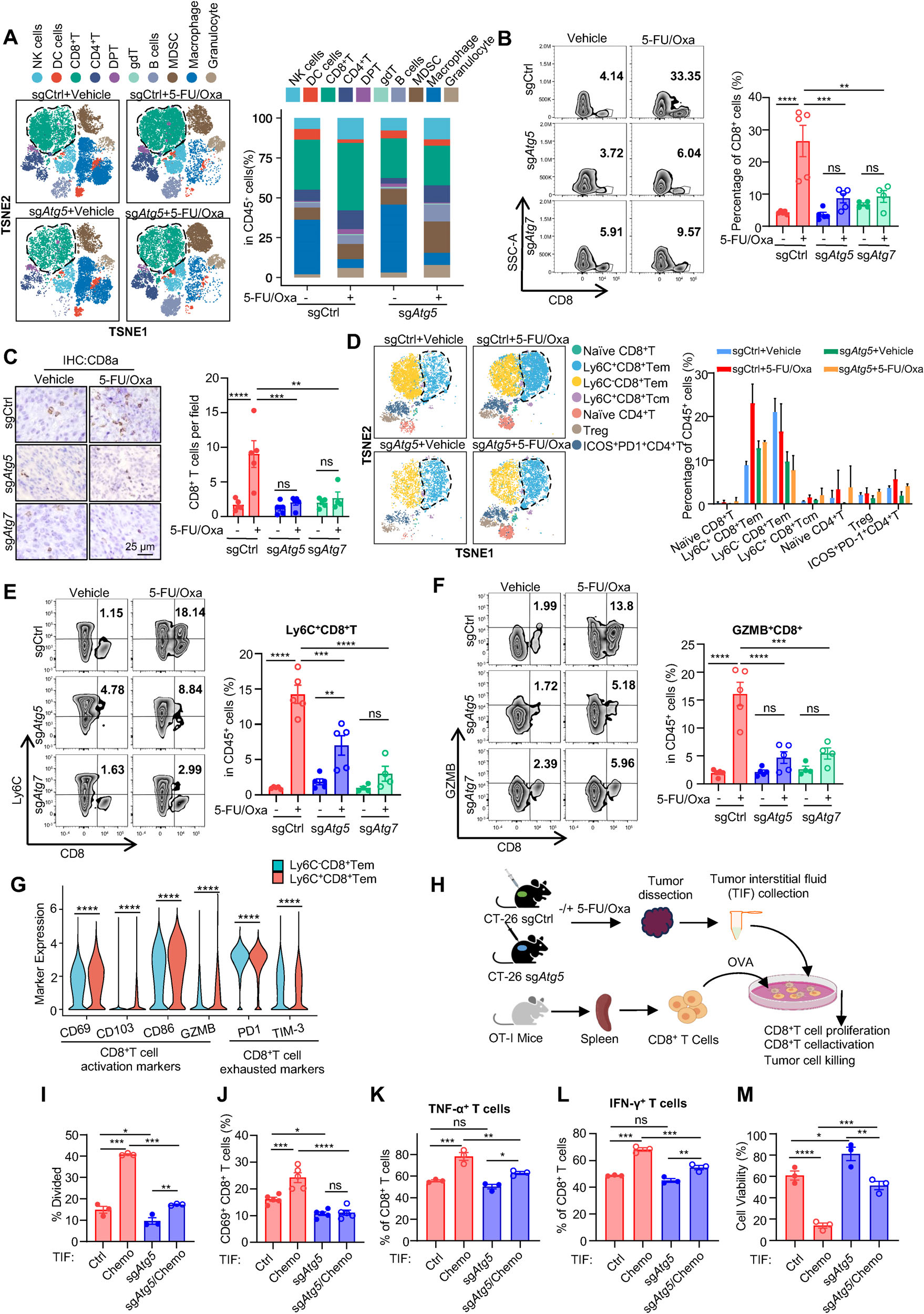
Fig. 2. Anti-tumor immunity evoked by chemotherapy depends on tumor cell-intrinsic autophagy. (A) t-SNE plots (left) and the frequency (right) of immune cell subsets in each treatment group, as determined by CyTOF (n = 2 per group). Each dot represents a single cell, with cell types color-coded. (B) Flow cytometric analysis and (C) immunohistochemical (IHC) staining of CD8+ T cells in sg Atg5 or sg Atg7 tumors following 5-FU/Oxa treatment (scale bar: 25 µm). (D) t-SNE plots (left) and quantification (right) of CD8+ and CD4+ T-cell subpopulations identified by CyTOF. (E,F) Flow cytometric analysis of activated CD8+ T cells (Ly6C+ or GZMB+ ) and quantitative summary of the indicated subsets. (G) Relative expression levels of activation and exhaustion markers in Ly6C+ versus Ly6C− CD8+ T cells, assessed by CyTOF. (H) Schematic showing of the in vitro co-culture system using spleen-derived CD8+ T cells and tumor interstitial fluid (TIF) from sgCtrl or sg Atg5 tumors. (I) CFSE staining analysis of CD8+ T cells proliferation(图注取自PDF文本层,来源:Advanced Science, 2026)
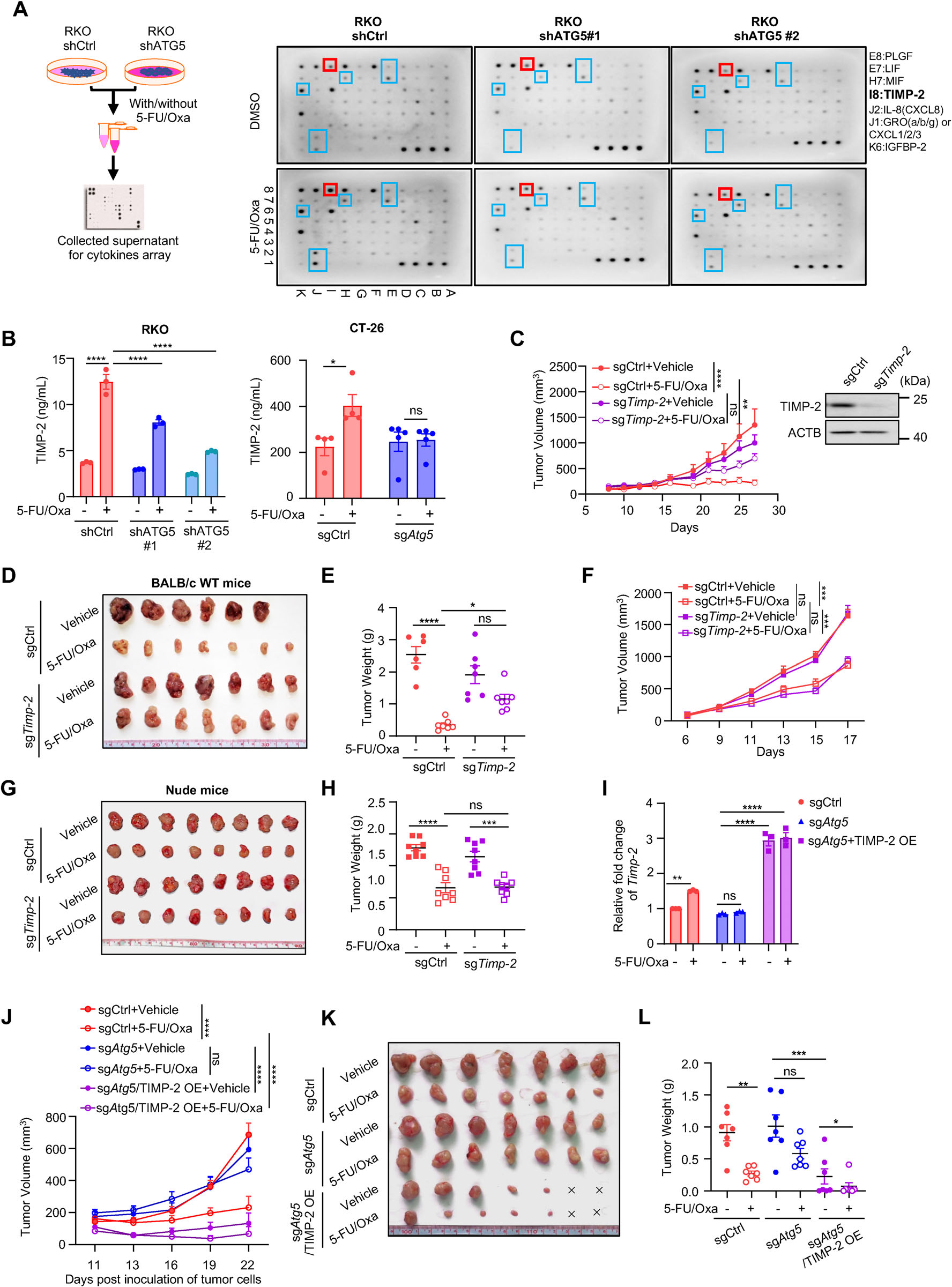
Fig. 3. TIMP-2 is a downstream effector of the autophagy-mediated response to chemotherapy. (A) Left: Schematic workflow for collecting cell culture supernatant from RKO cells expressing shCtrl or shATG5 after 48 h treatment with 5-FU/Oxa (100 µM/2.5 µM). Right: Membrane-based cytokine antibody array of secreted cytokines from the indicated groups; boxed spots indicate cytokines induced by 5-FU/Oxa in an autophagy-dependent manner. (B) ELISA quantification of TIMP-2 levels in culture supernatants from RKO and CT-26 cells with or without ATG5 silencing under 5-FU/Oxa treatment. (C–H) Growth curves of tumors, representative images, and tumor weights for CT-26 tumors with or without Timp-2 knockout(sgCtrl vs sg Timp-2) in WT (C–E) or nude (F–H) mice (n = 6-8 per group). Timp-2 knockout was confirmed by WB. (I) Generation of autophagy-deficient CT-26-sg Atg5 cells with or without ectopic TIMP-2 expression; TIMP-2 mRNA was confirmed by qRT-PCR. (J–L) Growth curves of tumors, representative images, and tumor weights for CT-26 tumors from sgCtrl or sg Atg5 cells with or without TIMP-2 overexpression (TIMP-2 OE), under the indicated treatments. Data are shown as mean ± SEM. Statistical analysis was performed using Two-way ANOVA with multiple comparisons. Significance is indicated as p < 0.05, p < 0.01, p < 0.001, p < 0.0001, ns, not significant.(图注取自PDF文本层,来源:Advanced Science, 2026)
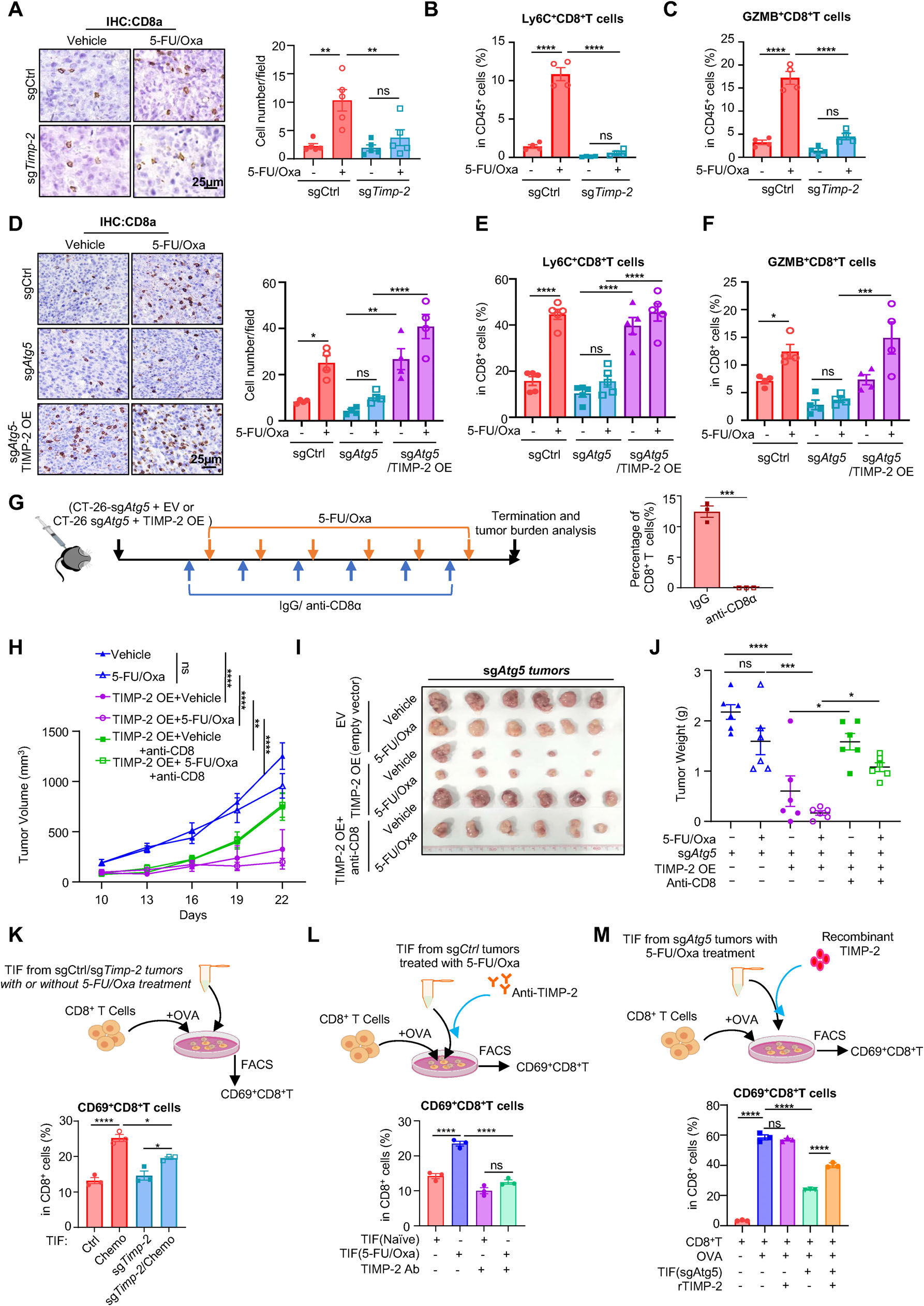
Fig. 4. TIMP-2 rescues chemotherapy sensitivity through activation of CD8+ T cells. (A) Representative IHC images(left) and quantifica- tion(right) of infiltrated CD8+ T cells in control or TIMP-2 knockout tumors treated with vehicle or 5-FU/Oxa (scale bar: 25 µm). (B,C) Flow cytometric quantification of activated CD8+ T cell subsets (Ly6C+ or GZMB+ ) in the same groups as (A). (D) Representative IHC images and quantification of CD8+ T cell infiltration in CT-26 sgCtrl, sg Atg5 , and sg Atg5 /TIMP-2 OE tumors under vehicle or 5-FU/Oxa treatment (scale bar: 25 µm). (E,F) Flow cytometric quantification of activated CD8+ T populations (Ly6C+ and GZMB+ ) for the groups in (D). (G) Schematic of the treatment protocol for CD8+(图注取自PDF文本层,来源:Advanced Science, 2026)
第四步:TIMP-2通过抑制MMP-2/9活性“唤醒”CD8⁺ T细胞
既然TIMP-2是MMP-2/9的抑制剂,那它的免疫调控功能是否依赖这个酶活抑制?
他们构建了两种丧失MMP抑制活性的TIMP-2突变体(Ala⁺突变体和N端截短突变体),在自噬缺陷细胞中分别回补野生型TIMP-2或两种突变体。结果只有野生型TIMP-2能恢复化疗敏感性和CD8⁺ T细胞浸润,两个突变体完全无效。
更直接的是,他们用MMP-2/9小分子抑制剂SB-3CT处理自噬缺陷小鼠,发现能显著恢复化疗疗效和CD8⁺ T细胞浸润。体外实验也证实,MMP-9会抑制OVA诱导的CD8⁺ T细胞活化,而重组TIMP-2能逆转这一效应。
@方法论点评:用“功能丧失突变体”来证明某个表型依赖于该蛋白的特定酶活功能,比单纯敲除更精准,能区分“蛋白本身存在”和“蛋白功能存在”两个层面。此外,用小分子抑制剂回补表型,也为临床转化提供了直接证据——如果自噬缺陷的病人,用MMP-2/9抑制剂能否“替补”TIMP-2的缺失?这个思路很有价值。
到这里,问题又反转了:自噬是怎么调控TIMP-2表达的?
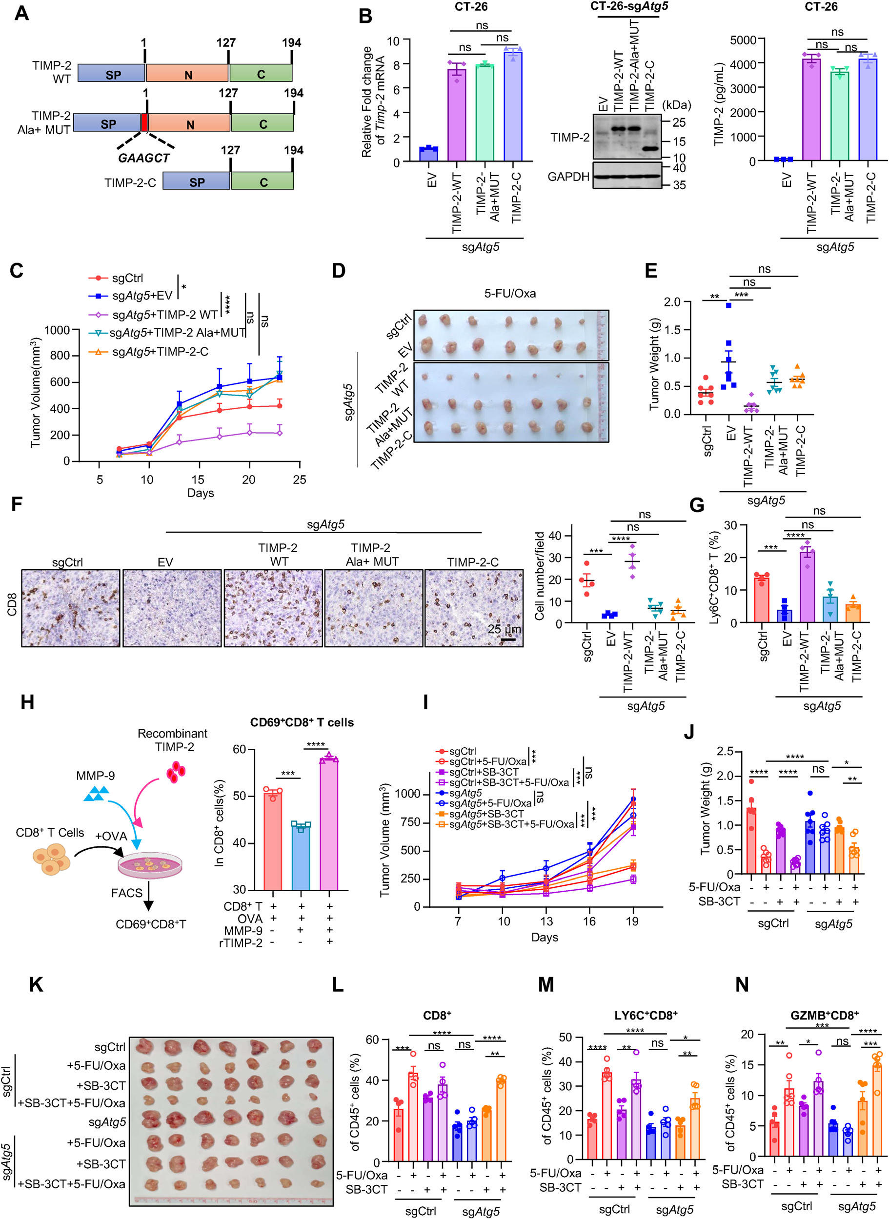
Fig. 5. Autophagy-TIMP-2 axis regulates anti-tumor immunity triggered by chemotherapy via modulating MMP-2/9 activity. (A) Schematic diagram showing TIMP-2 wild-type (WT) and its loss-of-function mutants, which are TIMP-2 mutant with an extra alanine—TIMP-2 Ala+ MUT and N terminal truncated mutant-TIMP-2-C. (B) The expression of TIMP-2 was confirmed by qRT-PCR, WB and ELISA. C) CT-26-sg Ctrl or sg Atg5 cells harboring TIMP-2 WT, TIMP-2 Ala+ MUT, TIMP-2-C or empty vector (EV) as control were inoculated into BALB/c WT mice and subjected to the 5- FU/Oxa treatment. Tumor growth curves were monitored over time. (D,E) Representative tumor images and quantitative analysis of tumor weights at sacrifice for the indicated groups (n = 7 per group). (F) Representative IHC images and quantification of intratumoral CD8+ T cell infiltration in the indicated groups as in (C) following 5-FU/Oxa treatment. Scale bar:25 µm. (G) FACS analysis of the proportions of active Ly6C+ CD8+ T cells in(图注取自PDF文本层,来源:Advanced Science, 2026)
第五步:自噬降解FBXW2→稳定C/EBPβ→转录激活TIMP-2
TIMP-2的mRNA水平受自噬调控,说明是转录水平的事件。他们用TMT蛋白质组学筛选自噬依赖性的差异蛋白,再叠加三个公开数据库预测TIMP-2的转录因子,结果交集只有一个——C/EBPβ。
验证很严谨:
敲低C/EBPβ,化疗诱导的TIMP-2上调被阻断;过表达C/EBPβ则促进TIMP-2转录。 双荧光素酶报告基因和ChIP实验证实C/EBPβ直接结合TIMP-2启动子上的两个位点(BS1和BS2)。 那么自噬如何影响C/EBPβ?质谱数据显示自噬调控了8个E3泛素连接酶,而FBXW2有最多的LIR基序(自噬受体结合基序),提示它可能直接被自噬降解。 机制:化疗促进FBXW2与LC3共定位(进入自噬体降解),导致FBXW2蛋白减少,从而解除对C/EBPβ的“压制”——FBXW2作为E3泛素连接酶,通过K48位点多泛素化C/EBPβ的K302和K316,将其导向蛋白酶体降解。
@方法论点评:这套“蛋白质组学筛选 + 公共数据库交集 + 分子互作验证”是经典的多层次机制挖掘路径。关键转折点在于:他们意识到自噬不仅是“降解”功能,还可以通过降解一个E3酶来“间接稳定”另一个蛋白——这是一种“去抑制”机制,而非直接的合成增加。这个逻辑在自噬研究中容易被忽略。
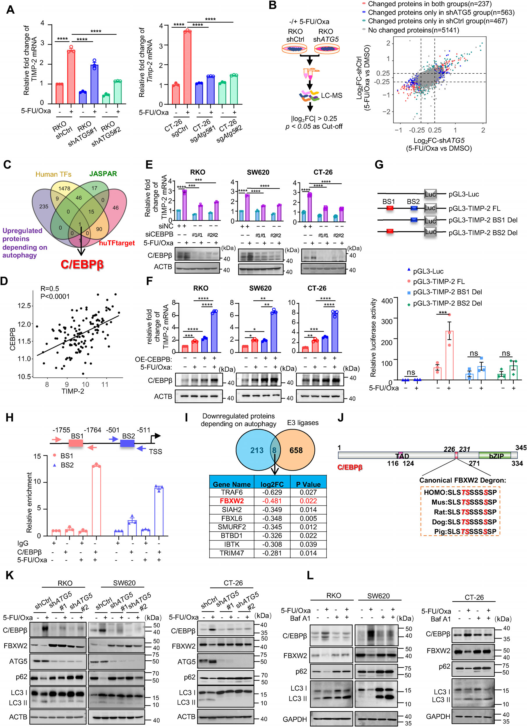
Fig. 6. FBXW2-C/EBP βaxis mediates autophagy-dependent TIMP-2 upregulation induced by chemotherapy. (A) qRT-PCR analysis of TIMP- 2 mRNA expression upon treatment with 5-FU/Oxa, with or without autophagy inhibition, in RKO and CT-26 cells. (B) Left: experimental timeline for collecting cell pellets for LC-MS analysis. Right: scatter plot of differentially expressed proteins (DEPs) between indicated groups. In the shCtrl group alone, a total of 467 DEPs were identified, comprising 246 upregulated and 221 downregulated proteins. (C) Venn diagram depicting the overlap of transcription factors derived from four datasets. (D) Correlation between CEBPB and TIMP-2 expression in CRC tissues (GSE72970). (E) qRT-PCR analysis of TIMP-2 mRNA in RKO, SW620, and CT-26 cells transfected with scrambled siRNA(si-Ctrl) or two independent siRNAs targeting CEBPB, with or without 5-FU/Oxa treatment. CEBPB knockdown efficiency was confirmed by WB. F) qRT-PCR analysis of TIMP-2 mRNA in RKO, SW620, and CT-26(图注取自PDF文本层,来源:Advanced Science, 2026)
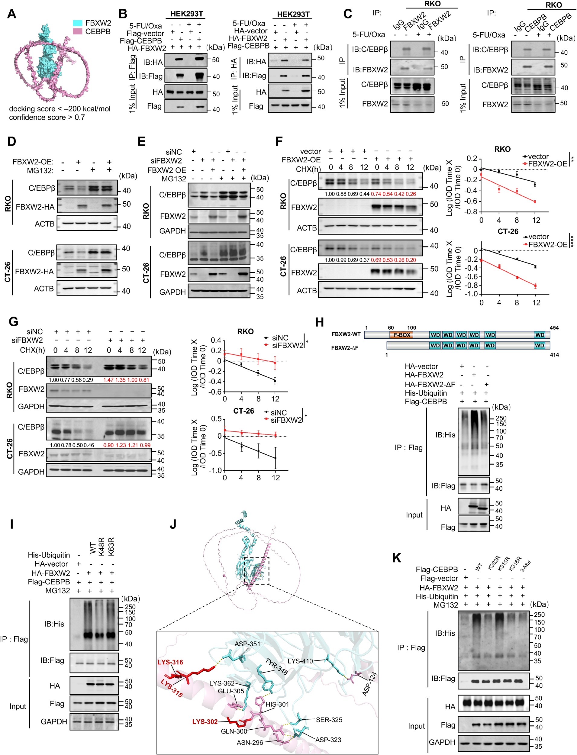
Fig. 7. FBXW2 interacts with C/EBP β and promotes its degradation through ubiquitin proteasome pathway. (A) The computational modeling of FBXW2 and C/EBP β interaction was performed by AlphaFold3, followed by visualization with PyMOL. Proteins color assignment: CEBPB (Purple), FBXW2 (Blue). (B) Exogenous interaction between C/EBP β and FBXW2 was examined by co-immunoprecipitation (Co-IP) in HEK293T cells transfected with the indicated plasmids, with or without 5-FU/Oxa treatment. Immunoprecipitation was performed with anti-Flag or anti-HA antibodies. (C) Co-IP analysis of endogenous interaction between C/EBP β and FBXW2 upon treatment with 5-FU/Oxa or not. Immunoprecipitation was performed using antibody against FBXW2 or C/EBP β. (D) Immunoblot analysis of C/EBP β expression in RKO and CT-26 cells overexpressing FBXW2, with or without proteasome inhibitor MG132 (20 µm, 6 h) treatment. (E) Immunoblot analysis of C/EBP β expression in RKO and CT-26 cells with FBXW2 knockdown(图注取自PDF文本层,来源:Advanced Science, 2026)
第六步:临床样本验证——自噬-TIMP-2轴与患者疗效正相关
最后回到临床:在多个公开CRC数据集中,自噬评分与TIMP-2表达正相关,TIMP-2高表达的患者对5-FU化疗更敏感,且预后更好。在他们自己的新辅助化疗队列中,应答者(TRG2)的肿瘤区域p-ATG16L1(自噬标志)和TIMP-2显著高表达,同时伴有更多GZMB⁺ T细胞浸润,而非应答者则相反。
@方法论点评:临床相关性的验证是整条链的“封顶”。机制做得再漂亮,如果在病人身上看不到相关性,转化价值就会打折扣。这里用TRG评分客观分层,并用多重免疫荧光共定位直接看到“自噬活化区域→TIMP-2高表达→T细胞活跃”的空间关联,增加了说服力。
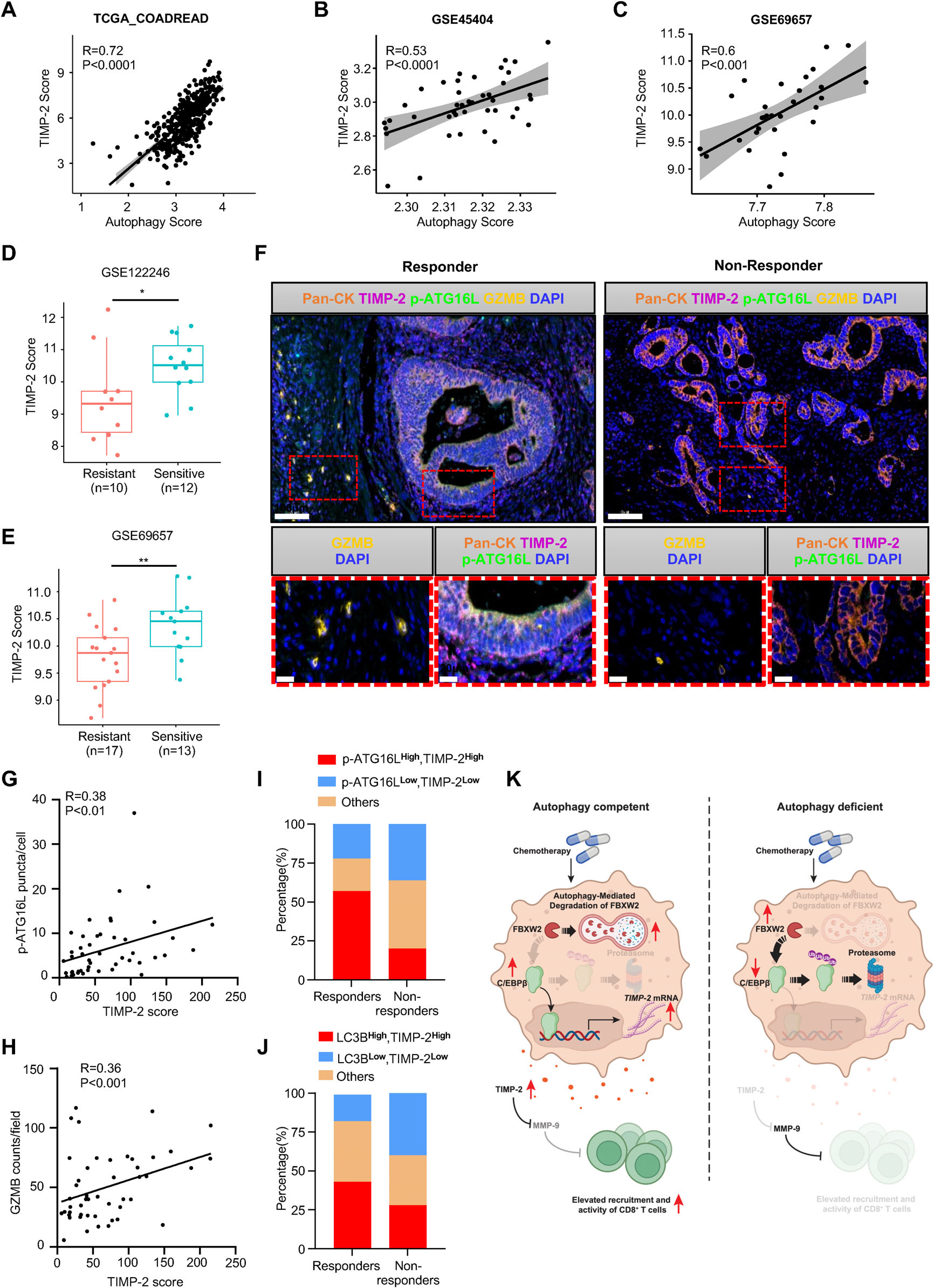
Fig. 8. Clinical relevance of the autophagy-TIMP-2 axis in CRC patients. (A–C) Correlation between TIMP-2 expression and autophagy score. (D,E) TIMP-2 expression levels in resistant versus sensitive CRC patients treated with 5-FU-based chemotherapy, derived from two independent cohorts. (F) Representative multiplex immunofluorescence images of responder and non-responder patient samples, co-stained for Pan-CK (orange), TIMP-2 (pink), p-ATG16L1 (green), and GZMB (gold). (G) Correlation between TIMP-2 expression and p-ATG16L1 puncta per cell, indicating autophagy activity, in the SYSU-the sixth Affiliated Hospital cohort (n = 48). (H) Correlation between TIMP-2 expression and GZMB counts per field, indicating active anti-tumor immunity, in the same cohort as in (G). (I) Patients were stratified into responders and non-responders based on the tumor regression grade (TRG) score, with TRG2 defined as responder (n = 23) and TRG3 (n = 25) as non-responder. Proportions of patients with p-ATG16L1High /TIMP-2High ; p-ATG16L1Low /TIMP-2Low or others were analyzed and shown in the graph. (J) Proportions of patients with high or low expression of LC3-II puncta, and others were analyzed and depicted in the graph, comparing responders and non-responders. (K) Schematic diagram illustrating tumor cell-intrinsic autophagy acts via the FBXW2/C/EBP β/TIMP-2 axis to regulate the TIME and chemosensitivity.(图注取自PDF文本层,来源:Advanced Science, 2026)
核心结论
肿瘤细胞内在的自噬通过FBXW2/C/EBPβ/TIMP-2轴,在化疗过程中重塑TIME,促进CD8⁺ T细胞浸润和活化,从而增强化疗敏感性。这条轴的核心逻辑是:
化疗激活自噬 → 自噬降解E3酶FBXW2 → FBXW2减少 → C/EBPβ蛋白稳定 → C/EBPβ转录激活TIMP-2 → TIMP-2抑制MMP-2/9 → MMP-2/9对CD8⁺ T细胞的抑制作用被解除 → CD8⁺ T细胞活化 → 肿瘤被清除。
这揭示了自噬在实体瘤化疗中的一个全新“正面”角色:它不仅是细胞的“清道夫”,更是免疫微环境的“外交官”——通过分泌性因子把免疫系统“叫来帮忙”。
对耐药/DTP/PGCC 的启示
自噬水平的异质性可能是化疗耐药的新分层标志:本研究显示自噬缺失的肿瘤在免疫完整环境下反而耐药,这提示某些DTP(药物耐受持久细胞)可能处于自噬活性低下状态,通过“免疫逃逸”而非“存活优势”来抵抗治疗。临床上或许需要区分“自噬依赖型耐药”和“自噬缺陷型免疫逃逸型耐药”。 TIMP-2/MMP-2/9轴是连接自噬和免疫的关键节点:对于自噬功能受损的耐药肿瘤,直接补充TIMP-2或使用MMP-2/9抑制剂(如SB-3CT)可能绕过自噬缺陷,恢复免疫应答。这为“靶向微环境”而非“靶向肿瘤细胞本身”的联合治疗策略提供了新思路。 PGCC(多倍体巨细胞)的自噬状态值得关注:PGCC常与化疗耐药和免疫抑制相关,但PGCC的自噬活性是否影响其免疫调控能力尚不清楚。本文的框架提示,检测PGCC中FBXW2/C/EBPβ/TIMP-2轴的活性,可能解释部分PGCC介导的免疫逃逸机制。
局限
模型系统:主要使用CT-26皮下移植瘤模型,虽然是免疫正常小鼠,但皮下微环境与肠道原位还是有差异;未在自发肿瘤或原位模型中全面验证。 TIMP-2的双重角色未完全排除:虽然证明了TIMP-2通过MMP-2/9起作用,但TIMP-2还有MMP非依赖的功能(如受体信号、细胞周期调控),这些是否也参与了免疫调控尚不能完全排除。 临床样本量偏小:自家队列n=48,需要更大规模验证;且未分析自噬-TIMP-2轴在MSI-H vs MSS亚型中的差异(本文主要用MSS模型)。 FBXW2的底物不止C/EBPβ:FBXW2还有其他已知底物(如β-catenin、AKT),这些分子也可能参与了免疫调控,文中未深入探讨它们是否协同贡献。
来源
期刊:Advanced Science,2026年。DOI:10.1002/advs.77349