一句话亮点
本文发现DIXDC1通过直接结合并稳定代谢酶DLAT,用一个“双头钉”机制同时推动膀胱癌的淋巴转移和抵抗铜死亡,揭示了代谢适应性背后的隐藏调控者。
背景/痛点
膀胱癌一旦发生淋巴转移,五年生存率直接从70%掉到30%,是个硬骨头。肿瘤转移需要强大的能量支撑,往往要依赖线粒体氧化磷酸化和三羧酸循环(TCA)。但这里有个“搬起石头砸自己脚”的悖论:TCA循环的关键酶——同时也是铜死亡的核心执行者DLAT——在铜离子过载时会发生毒性寡聚化,直接把细胞送上西天。
那转移能力强的癌细胞是怎么既利用DLAT产能,又避免被铜死亡干掉的?这就是本文要解决的核心矛盾。作者把目光投向了DIXDC1,一个经典的Wnt信号通路支架蛋白,但他们怀疑这家伙还有别的“副业”。
推理链分步拆解
第一步:先看临床相关性——DIXDC1是不是找对了对象?
作者首先在两个独立临床队列里做了IHC染色,发现DIXDC1在膀胱癌组织里明显高表达,而且在淋巴结阳性、肌层浸润性、高级别肿瘤里更高。TCGA数据库的mRNA数据也呼应了这一趋势。Kaplan-Meier曲线显示DIXDC1高表达的患者总生存期和无病生存期都更短。
@方法论点评:临床相关性是机制探索的“合法入场券”。如果没有这一步,后面做得再漂亮也是空中楼阁。多队列、多数据库验证是肿瘤研究中避免“单中心偶然性”的基本操作。
接着用脚垫注射模型评估体内淋巴转移能力,结果DIXDC1过表达组腘窝淋巴结明显肿大,GFP阳性肿瘤细胞浸润增加——直接证明DIXDC1是淋巴转移的推手。他们选脚垫模型是因为足爪的淋巴引流路径清晰,便于定量评估淋巴转移。
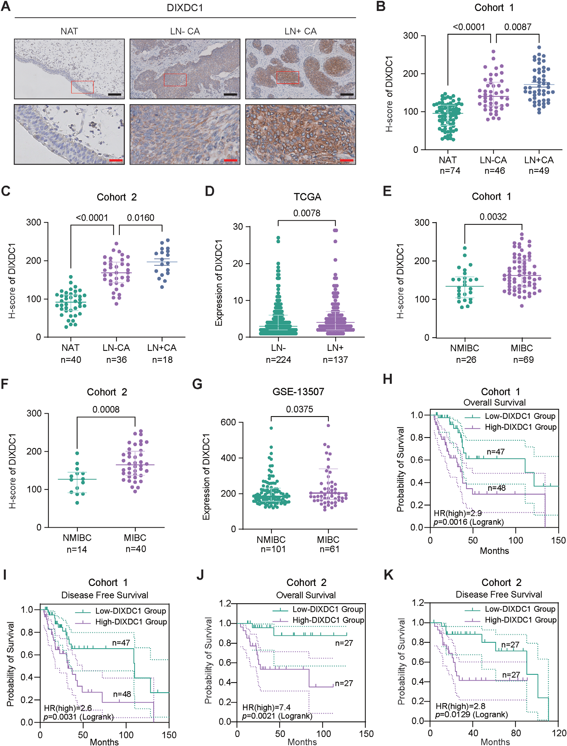
Fig. 1. DIXDC1 is overexpressed and associated with poor prognosis in bladder cancer. (A) Representative immunohistochemical (IHC) staining images showing DIXDC1 expression in adjacent normal tissues (NAT), lymph node-negative bladder cancer tissues (LN− CA), and lymph node-positive bladder cancer tissues (LN+ CA), with high-magnification images of the red-boxed regions shown below. Scale bar: 100 µm (black), 20 µm (red). (B, C) Quantitative H-score analysis of DIXDC1 protein expression in NAT, LN− CA, and LN+ CA tissues from Cohort 1 (B) at the Second Affiliated Hospital of Kunming Medical University and Cohort 2 (C) at Sun Yat-sen Memorial Hospital. (D) Analysis of DIXDC1 mRNA expression levels in lymph node-negative and lymph node-positive bladder cancer samples based on data from The Cancer Genome Atlas (TCGA). (E, F) Comparative analysis of DIXDC1 protein expression between non-muscle-invasive bladder cancer (NMIBC) and muscle-invasive bladder cancer (MIBC) tissues in Cohort 1 (E)(图注取自PDF文本层,来源:Advanced Science, 2026)
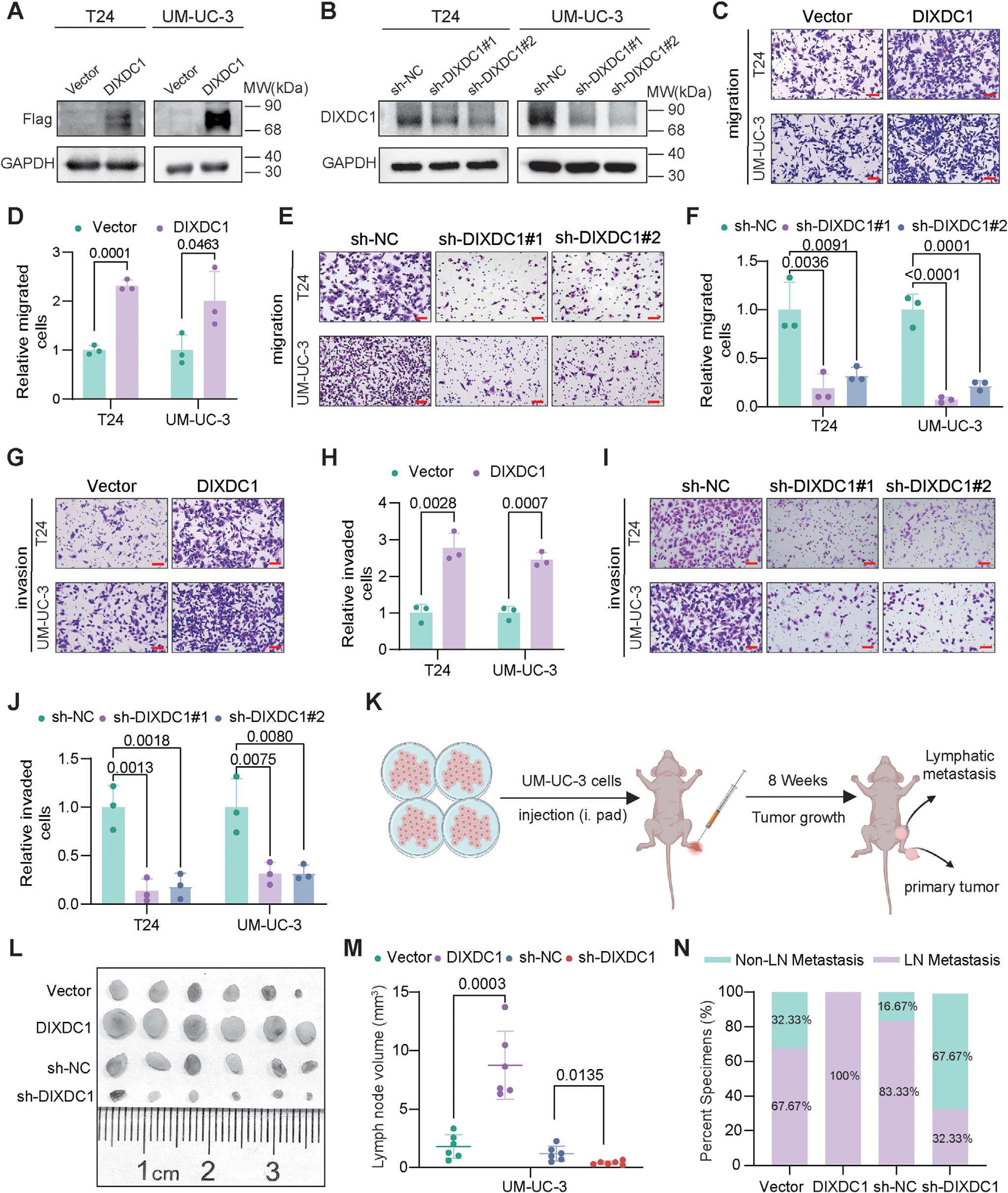
Fig. 2. DIXDC1 promotes the metastasis-related behaviors of bladder cancer cells in vitro and enhances lymph node metastasis in vivo. (A) Verification of DIXDC1 overexpression in T24 and UM-UC-3 cells transfected with either an empty vector or a DIXDC1 expression construct by Western blot. (B) Assessment of DIXDC1 protein expression in T24 and UM-UC-3 cells with DIXDC1 silencing, determined by Western blot. (C, D) Representative images (C) and quantitative analysis of migrated cell counts (D) from Transwell migration assays in T24 and UM-UC-3 cells overexpressing DIXDC1. (E, F) Representative images (E) and quantitative analysis of migrated cell counts (F) in DIXDC1 silencing. (G, H) Representative images (G) and quantitative analysis of invaded cell counts (H) in overexpressing DIXDC1. (I, J) Representative images (I) and quantitative analysis of invaded cell counts (J) in DIXDC1 silencing. Scale bar: 200 µm (red). (K) Schematic diagram illustrating the experimental workflow for establishing a murine plantar footpad injection/lymph node metastasis model with popliteal lymph node metastasis using UM-UC-3 cells. (L) Representative gross anatomical images(图注取自PDF文本层,来源:Advanced Science, 2026)
第二步:找靶点——DIXDC1到底“抱住”了谁?
那DIXDC1是通过什么机制干活的?既然它是支架蛋白,最可能的方式是直接结合某个“关键效应蛋白”。他们用Co-IP银染结合质谱,在55-70 kDa附近钓到了一个差异条带——DLAT。随后用内源性Co-IP、免疫荧光共定位和分子对接进一步验证,确认了DIXDC1与DLAT的物理结合。
这里有个关键设计:他们构建了DIXDC1的一系列截短突变体,发现缺失600-680 aa区域(C端DIX结构域)后结合能力下降最明显,说明这个区域是关键结合界面。
@方法论点评:截短突变体是“哪个部位干活”的黄金标准,比单纯敲低/过表达更精细。此外,分子对接+实验验证的组合也是结构生物学与细胞生物学联动的常见套路。

Fig. 3. DIXDC1 binds and stabilizes DLAT by inhibiting its ubiquitin-mediated degradation. (A) Silver staining analysis following co- immunoprecipitation of endogenous DIXDC1 in T24 and UM-UC-3 cells, with DIXDC1-interacting proteins identified by mass spectrometry; the red arrows indicate the bands corresponding to DIXDC1 and DLAT. (B) In silico prediction of the DIXDC1-DLAT interaction and their potential binding interface. Structural domains of DIXDC1 (yellow) and DLAT (pink) were retrieved from the Protein Data Bank (PDB). Rigid protein-protein docking was performed using the Gram-X web server, and the predicted DIXDC1-DLAT interaction and binding interface were visualized using PyMOL. (C) Co-immunoprecipitation assays confirming the endogenous protein-protein interaction between DIXDC1 and DLAT in T24 and UM-UC- 3 cells. (D) Confocal immunofluorescence microscopy showing the subcellular colocalization of DIXDC1 and DLAT. Scale bar: 10 µm (white). (E, F) Schematic representation of DIXDC1 deletion mutants used to evaluate the contribution of different DIXDC1 regions to DLAT binding (E), and(图注取自PDF文本层,来源:Advanced Science, 2026)
第三步:稳定DLAT——DIXDC1是怎么帮DLAT“续命”的?
接下来要回答:DIXDC1结合DLAT之后发生了什么?他们发现DIXDC1过表达或敲低能平行改变DLAT蛋白水平,但不影响DLAT的mRNA水平,说明调控发生在蛋白层面,而不是转录层面。CHX放线菌酮追踪实验显示DIXDC1敲低后DLAT降解速度加快;而泛素化实验(MG132处理 + HA-Ub)证明DIXDC1敲低增加了DLAT的多聚泛素化水平。
@方法论点评:区分mRNA和蛋白水平变化是判断“调控层级”的关键一步。这里排除了转录调控,把因果链锁定在“蛋白稳定性/降解”上。放线菌酮实验则是评估蛋白半衰期变化的经典工具——如果敲低后蛋白降解加快,说明稳定效应消失了。
第四步:下游信号——DLAT被稳住之后,又干了什么?
DLAT是代谢酶,但作者在RNA-seq中发现DIXDC1敲低会影响大量基因的转录水平。经过交叉筛选,他们锁定了NEK7和CCNE2——这两个基因的表达在DIXDC1和DLAT敲低后都显著下降,而且DIXDC1过表达上调它们、DIXDC1敲低下调它们,这个调控依赖DLAT(DLAT敲低可以部分取消DIXDC1过表达引起的上调)。
关键是机制层面:放线菌素D chase实验显示DIXDC1敲低加速了NEK7和CCNE2 mRNA的降解,而过表达则延缓降解——DLAT是通过增强mRNA稳定性来调控它们的,而不是通过转录激活。那DLAT作为一个线粒体代谢酶,是怎么跑到胞质里去调控mRNA稳定性的?文中没说清楚,但这正是“代谢酶的非经典功能”(moonlighting function)的有趣之处——类似PKM2、PHGDH已被报道过有RNA调控功能。
功能验证中,回补NEK7或CCNE2能够部分挽救DIXDC1敲低引起的迁移和侵袭缺陷,说明这俩确实是下游功能执行者。
@方法论点评:交叉验证(DIXDC1敲低 vs DLAT敲低)是区分“直接靶点”与“伴随效应”的经典过滤策略。如果两个不同操作能筛选出交集基因,那这些基因更可能是同一通路的核心节点。
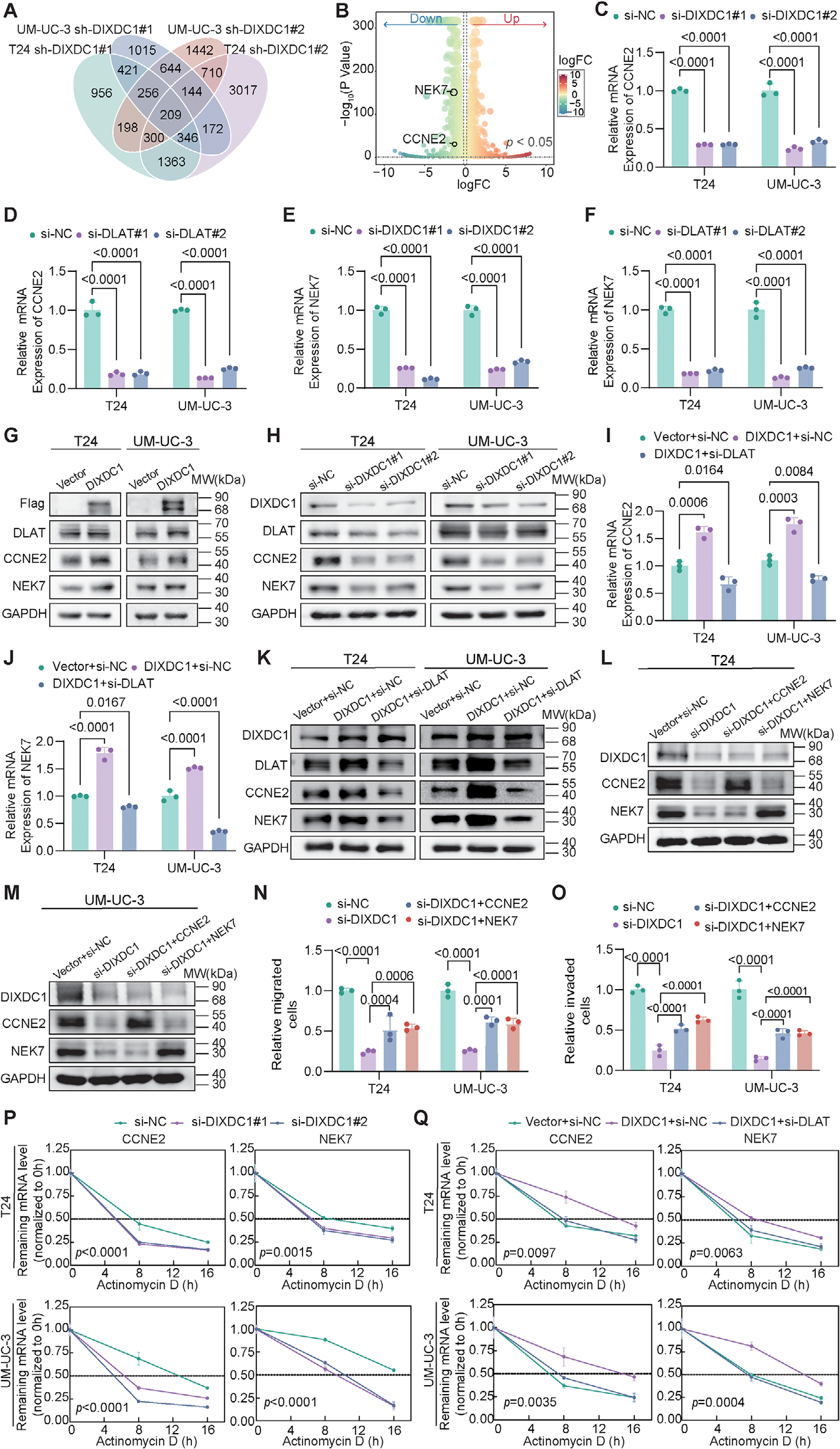
Fig. 4. DIXDC1 regulates the expression and mRNA stability of NEK7 and CCNE2 via DLAT. (A) Transcriptome sequencing analysis of T24 and UM-UC-3 cells with DIXDC1 knockdown to identify differentially expressed genes (DEGs), with DEG criteria defined as |log2 fold change| ≥ 0.585, corresponding to a 1.5-fold change, and adjusted p value (FDR) < 0.05. (B) Volcano plot of DEGs following DIXDC1 knockdown, with the downregulated candidate genes CCNE2 and NEK7 highlighted. (C–F) qRT-PCR quantification of relative mRNA expression of CCNE2 and NEK7 in T24 and UM-UC-3 cells. Transcript levels of CCNE2 and NEK7 following transfection with si-NC or two separate DIXDC1-targeting siRNAs (C, E); transcript levels of CCNE2 and NEK7 following transfection with si-NC or two separate DLAT-targeting siRNAs (D, F). (G, H) Western blot analysis was conducted to determine the protein levels of DIXDC1, DLAT, NEK7, and CCNE2 in T24 and UM-UC-3 cells under conditions of DIXDC1 overexpression (G) or knockdown (H). (I, J) qRT-PCR quantification of CCNE2 (I) and NEK7 (J) mRNA expression in T24 and UM-UC-3 cells with DIXDC1 overexpression following subsequent(图注取自PDF文本层,来源:Advanced Science, 2026)

Fig. 5. DIXDC1 promotes bladder cancer cell proliferation in vitro. (A, B) Cell viability assays at multiple time points using CCK-8 and growth curve generation in DIXDC1-overexpressing T24 (A) and UM-UC-3 (B) cells. (C, D) Colony formation assay in DIXDC1-overexpressing T24 and UM-UC-3 cells, with representative colony images (C) and corresponding quantitative analysis of colony formation efficiency (D). (E, F) Cell viability measurement at different time points by CCK-8 and growth curve plotting in T24 (E) and UM-UC-3 (F) cells with DIXDC1 knockdown. (G, H) Colony formation assay in T24 and UM-UC-3 cells with DIXDC1 knockdown, with representative colony images (G) and corresponding quantitative analysis of colony numbers (H). (I, J) In T24 (I) and UM-UC-3 (J) cells with stable overexpression of DIXDC1, DLAT was further knocked down, and cell growth curves were evaluated using the CCK-8 assay. (K, L) Colony formation assay under the indicated experimental conditions, with representative colony images (K) and corresponding quantitative analysis of colony numbers (L). Data are presented as means ± standard deviations (SD). Statistical analyses were performed using two-way ANOVA followed by Šídák’s multiple-comparisons test for panels (A, B), two-way ANOVA followed by Dunnett’s multiple-comparisons test for panels (E, F, I, J), unpaired Student’s t -test for panel (D), and one-way ANOVA followed by Dunnett’s multiple-comparisons test for panels (H, L).(图注取自PDF文本层,来源:Advanced Science, 2026)
第五步:铜死亡——DIXDC1怎么让细胞“不怕铜”了?
既然DLAT是铜死亡的核心执行者,那DIXDC1结合并稳定DLAT,会不会也调节铜死亡敏感性?这是本研究的亮点之一。
他们首先测量了膀胱癌组织中的铜离子浓度,发现显著高于正常组织。体外实验中,DIXDC1敲低降低了ES-Cu(铜离子载体+Cu²⁺)的IC₅₀,而过表达则提高了IC₅₀,说明DIXDC1让细胞更“抗铜死亡”。Ghost Dye流式细胞术进一步确认——DIXDC1敲低后ES-Cu诱导的细胞死亡增加,而且这种死亡能被铜螯合剂TTM逆转,说明是铜依赖性的细胞死亡,不是其他类型细胞死亡(凋亡、坏死、焦亡、铁死亡)的混淆效应。
更深层的机制证据在于:DIXDC1敲低后,脂酰化DLAT(Lip-DLAT)水平升高、DLAT寡聚化增加(非还原WB检测)。也就是说DIXDC1在稳定DLAT的同时,也抑制了它在铜刺激下的毒性寡聚化——这解释了为什么DIXDC1高表达的细胞既能利用DLAT的代谢功能,又不被铜死亡干掉。
@方法论点评:从“IC₅₀”到“死亡类型排除”再到“分子标志物(脂酰化+寡聚化)”是一套完整的铜死亡研究逻辑链。使用多种死亡通路抑制剂(Z-VAD、Nec-1、VX-765、Lip-1)排除其他死亡形式,就是为了防止读者质疑:这真的是铜死亡,不是其他类型细胞死亡?这一步虽然笨,但恰恰是最严谨的科研做法。

Fig. 6. DIXDC1 promotes resistance to copper-induced cuproptosis in bladder cancer. (A) Measurement of copper ion concentration in paired bladder cancer tissues and their corresponding adjacent normal tissues using a copper ion detection kit, with each connecting line representing an individual patient. (B) The copper ion content was measured in the normal uroepithelial cell line SV-HUC-1 and bladder cancer cell lines (5637, SW-780, T24, UM-UC-3, RT-112, HT-1376, RT-4) using a commercial copper ion detection kit. (C) Determination of the half-maximal inhibitory concentration (IC50 ) of the copper ionophore Elesclomol combined with Cu (II) (ES-Cu) in T24 and UM-UC-3 cells with DIXDC1 knockdown. (D) Measurement of ES-Cu IC50 in DIXDC1-overexpressing T24 and UM-UC-3 cells. (E, F) Ghost dye staining followed by flow cytometry analysis of cell death in T24 (E) and UM-UC-3 (F) cells under the indicated treatments. Quantification of Ghost dye–positive cells is shown in control and DIXDC1 knockdown cells treated with DMSO, TTM, ES-Cu, or ES-Cu + TTM. (G, H) Co-immunoprecipitation analysis of lipoylated DLAT (Lip-DLAT) and total DLAT in T24 (G) and(图注取自PDF文本层,来源:Advanced Science, 2026)
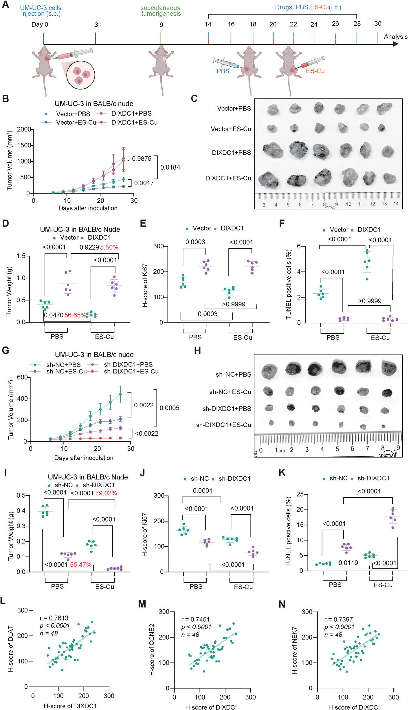
Fig. 7. DIXDC1 modulates the growth of bladder cancer cells and resistance to cuproptosis in vivo. (A) Schematic illustration of experimental design. Subcutaneous xenograft models were established in nude mice by inoculating UM-UC-3 cells with stable DIXDC1 overexpression or knockdown (and their respective controls). After tumor formation, mice were treated with PBS or ES-Cu (6 mg/kg, intraperitoneal injection, every 3 days). (B–F) Tumor volume (B), gross morphology (C), final tumor weight (D), Ki67 H-scores (E), and TUNEL positivity (F) were assessed in xenografts derived from UM-UC-3 cells with stable DIXDC1 overexpression. (G–K) Corresponding analyses for tumor volume (G), gross morphology (H), final tumor weight (I), Ki67 H-scores (J), and TUNEL positivity (K) were performed in xenografts with stable DIXDC1 knockdown. (L–N) Correlation analysis between DIXDC1 expression and the expression of DLAT (L), CCNE2 (M), and NEK7 (N) in subcutaneous xenograft tumors, based on H-score quantification. All data are presented as means ± standard deviations (SD). Statistical analyses were performed using two-way ANOVA followed by Tukey’s multiple-comparisons test for panels (B, D–G, I–K), and Pearson correlation analysis for panels (L, M).(图注取自PDF文本层,来源:Advanced Science, 2026)
第六步:体内验证——能不能在活体里复现?
皮下异种移植模型中,DIXDC1过表达促进肿瘤生长、削弱ES-Cu疗效;而DIXDC1敲低抑制肿瘤生长、增强ES-Cu疗效。Ki67和TUNEL染色分别证实了增殖和凋亡变化。
原位膀胱癌模型中,DIXDC1过表达增加膀胱肿瘤体积和重量,并且促进盆腔淋巴结转移;反之,DIXDC1敲低则减弱这一切。最后,DLAT敲低可以部分取消DIXDC1过表达带来的生长优势——证明DLAT是DIXDC1体内功能的关键下游。
@方法论点评:皮下模型看“生长+治疗响应”,原位模型看“器官特异性转移”,两者互补——皮下模型方便量化药物响应,原位模型更贴近临床。体内“rescue实验”是验证信号轴整体逻辑的最后一块拼图。

Fig. 8. DIXDC1 drives bladder cancer progression and lymphatic metastasis through DLAT in vivo. (A-F) Orthotopic bladder cancer xenograft models were established by inoculating luciferase-labeled stable DIXDC1-overexpressing or DIXDC1-knockdown human bladder cancer UM-UC-3 cells (and their respective control cells) into 4-5-week-old female BALB/c nude mice. (A) Representative in vivo bioluminescence imaging of tumor burden at the experimental endpoint (day 14 post-inoculation). (B) Representative images of orthotopic bladder tumors and pelvic lymph nodes. (C) Quantification of bladder tumor volume. (D) Measurement of bladder tumor weight. (E) Quantification of pelvic lymph node volume. (F) Western blot analysis confirming DIXDC1 overexpression with subsequent DLAT knockdown in the established rescue stable cell lines. (G–I) Subcutaneous xenograft models were established by inoculating the stable cell lines generated in (F) into 4–5 week old male BALB/c nude mice. (G) Representative images of excised subcutaneous tumors. (H) Tumor growth curves. (I) Final tumor weight at the experimental endpoint. (J) Schematic model illustrating the role of the DIXDC1-DLAT axis in bladder cancer progression and cuproptosis resistance. DIXDC1 directly interacts with DLAT, exerting a dual regulatory function. On the left, DIXDC1 stabilizes DLAT by inhibiting its ubiquitination and proteasomal degradation. The stabilized DLAT then enhances the(图注取自PDF文本层,来源:Advanced Science, 2026)
核心结论
DIXDC1通过直接结合DLAT,发挥“双面调控”功能:
一面促转移:抑制DLAT泛素化降解→稳定DLAT→增强NEK7/CCNE2 mRNA稳定性→促进细胞增殖、迁移、侵袭和淋巴转移; 一面抗死亡:限制DLAT脂酰化及铜诱导的毒性寡聚化→抵抗铜死亡。
这两个功能协同,让肿瘤细胞在“高速转移”和“存活”之间找到平衡。从科研角度,本文把代谢酶的非经典功能(mRNA稳定性调控)和新型细胞死亡(铜死亡)串联起来,是一个相当漂亮的机制故事。
对耐药/DTP/PGCC 的启示
铜死亡抵抗可能是DTP细胞存活的潜在机制:DTP(药物耐受持久细胞)往往依赖氧化磷酸化存活,而铜死亡恰恰攻击TCA循环。本文发现DIXDC1-DLAT轴能抵抗铜死亡,暗示DTP可能通过上调该轴来维持存活。可以在DTP模型中检测DIXDC1/DLAT表达,并探索诱导铜死亡是否能清除DTP。 传统化疗与铜死亡诱导剂的“合成致死”可能:肿瘤高表达DIXDC1意味着对铜死亡不敏感,但如果联合靶向DIXDC1或DLAT的抑制剂(如siRNA或小分子),有可能将耐药细胞“去耐受化”。文中体内实验也显示了ES-Cu在DIXDC1敲低时疗效增强。 PGCC(多倍体巨细胞)与铜死亡的关系值得探索:PGCC在化疗后出现,具有更强的代谢适应性和存活能力,本文发现的DIXDC1-DLAT轴可能在这些细胞中同样活跃。如果能在PGCC模型中验证,可能会开辟一条“诱导铜死亡清除PGCC”的新方向。
局限
功能验证主要依赖敲低和过表达,缺乏CRISPR/Cas9完全敲除的遗传学证据。虽然逻辑清晰,但敲低可能存在脱靶或残留,结论强度不如敲除。 DLAT如何增强NEK7/CCNE2 mRNA稳定性具体机制未阐明:是直接结合RNA,还是通过代谢物影响RNA结合蛋白?这需要进一步生化证据。 体内实验主要基于细胞系异种移植模型,未使用患者来源的异种移植(PDX)或类器官验证临床相关性和个体差异。 未系统评估DIXDC1在其他类型细胞死亡(如凋亡、铁死亡)中的潜在调节作用,可能存在更广泛的“存活卫士”功能。
来源
期刊:Advanced Science,2026。DOI: 10.1002/advs.77435