一句话亮点
这篇 2026 年的《Cell Death & Differentiation》文章发现,原本作为抗氧化系统的硒蛋白 TrxR1,在铁死亡中反而扮演"加速器"角色——它通过稳定 KEAP1、抑制 NRF2、降低 GPX4 表达,让癌细胞对铁死亡更敏感。更关键的是,这个机制依赖于 TrxR1 C 末端的硒代半胱氨酸 U498,而且还提供了用沙利度胺联合铁死亡诱导剂 IKE 协同抗癌的潜在策略。
背景/痛点
铁死亡的核心是脂质过氧化物积累到致死水平,而 GPX4 是这个过程的"守门人"——它负责把脂质过氧化物还原掉。GPX4 一失灵,细胞就死了。细胞内抗氧化系统主要有两套:GSH-GPX4 系统和 Trx-TrxR 系统。前者和铁死亡的关系已经研究得很清楚了,但后者在铁死亡中到底扮演什么角色,之前一直有争议。早前有报道说 TrxR1 是抑制铁死亡的,但这篇文章的作者觉得事情没那么简单——他们决定自己搞清楚。
推理链分步拆解
第一步:先确认 TrxR1 到底促进还是抑制铁死亡
作者之前做过一个 RNAi 筛选,意外发现 TrxR1 是个候选的"正调控因子"——这个结果跟他们自己预期的方向相反,于是他们决定从头验证。
他们先用 TrxR1 的特异性抑制剂 TRi-1 处理多种癌细胞(HT1080、MDA-MB-231、MEF、B16-F10),结果发现 TRi-1 居然能阻断 erastin 诱导的脂质过氧化和细胞死亡。而且 TRi-1 本身没有抗氧化活性(DPPH 法验证过),说明这个阻断效应不是因为它"顺便把自由基清了"。反过来,敲低 TrxR1 让细胞对 erastin 和 GPX4 直接抑制剂 RSL3 都变得不敏感;而过表达 TrxR1 则显著增强了脂质过氧化和铁死亡。
这里有个细节很有意思:erastin 处理会让 TrxR1 发生时间依赖性的二聚化,而二聚化对 TrxR1 发挥功能很重要。他们做了一个 W114R 突变破坏二聚化,结果这个突变体完全救不回 TrxR1 敲低细胞的铁死亡敏感性。所以,TrxR1 促进铁死亡这件事,功能上是成立的,而且需要二聚化。
@方法论点评:用抑制剂、敲低、过表达"三向验证"一个基因的功能,是分子生物学的基本操作。更关键的是他们排除了 TRi-1 的脱靶抗氧化效应(DPPH 实验),这种"排除混淆因素"的思维很重要。

Fig. 1. TrxR1 Is a Positive Regulator of Ferroptosis. A The TrxR1 inhibitor TRi-1 blocks erastin-induced lipid peroxidation and ferroptosis. Cells were treated with erastin (HT1080: 10 μΜ; MDA-MB-231: 15 μΜ; MEF: 5μM; B16-F10: 15μM) with or without TRi-1 for 12 h, cells were then stained with Propidium Iodide (PI) and imaged using fluorescence microscopy at 200 × magnification or analyzed by flow cytometry. Cells treated as indicated were stained with BODIPY 581/591 C11, and lipid ROS levels were measured by flow cytometry. Scale bar: 100 μm. B TRi-1 has no antioxidant activity measured by the DPPH assay. C Knockdown of TrxR1 inhibits erastin-induced lipid peroxidation and ferroptosis. Western blot images show the expression of the indicated protein. Cells as indicated were treated with erastin (HT1080: 10 μΜ; MDA-MB-231: 15 μM; B16-F10: 15μM) for 12 h or (MEF: 5 μM) for 6 h, then cell death and lipid ROS were measured as described in Fig. 1A. Scale bar: 100 μm. D Overexpression of TrxR1 promotes lipid peroxidation and ferroptosis. Western blot images and quantitative data confirm the expression of the indicated protein. Cells as indicated were treated with erastin (HT1080: 10 μΜ; MDA-MB-231: 15 μM; ACHN: 15 μM and B16-F10: 15 μM) for 8 h, then cell death and lipid ROS were measured as described in Fig. 1A. Fer-1 (2 μM), DFO (100 μM), Z-VAD-FMK (50 μM), NEC-2 (50 μM). Scale bar: 100 μm. E Knockdown of TrxR1 suppresses RSL3-induced lipid peroxidation and ferroptosis. TrxR1 KD HT1080 cells were treated with RSL3 as indicated for 10 h, then cell death and lipid ROS were measured as Fig. 1A. Scale bar: 100 μm. F Time-dependent dimerization of TrxR1 induced by erastin (10 μΜ). Western blot images and quantitative data confirm the expression of indicated protein. G The Tryptophan114th to Arginine114th (W114R) mutation of TrxR1 disrupts its dimerization. Western blot images and quantitative data show the expression of the indicated protein. H Dimerization of TrxR1 is required for its function in ferroptosis. HT1080 cells transfected with wild-type or TrxR1W114R were treated with erastin (10 μΜ) for 12 h, then cell death and lipid ROS were measured as Fig. 1A. Scale bar: 100 μm. Data are derived from three independent experiments, and each value represents the mean ± SD. P < 0.05, P < 0.01, P < 0.001, t test. (n = 3).(图注取自PDF文本层,来源:Cell Death & Differentiation, 2026)
第二步:那么 TrxR1 到底是通过什么机制促进铁死亡的?
既然表型确定了,下一步自然是找靶点。作者把 TrxR1 敲低细胞的蛋白裂解液跑了一遍 Western blot,发现 GPX4 和 FSP1 都上调了。GPX4 是铁死亡的核心负调控因子,这个上调可以解释为何细胞变耐受了。
奇怪的是,ACSL4(铁死亡的正调控因子)也上调了——按理说这会让细胞更敏感才对,但 TrxR1 敲低细胞反而耐药。作者判断 GPX4 和 FSP1 的上调盖过了 ACSL4 的作用。后续验证也支持这个判断:敲低 FSP1 能部分恢复 TrxR1 敲低细胞的铁死亡敏感性,说明 FSP1 确实也参与了,但主要矛盾还是 GPX4。
更有意思的是反向验证:过表达 TrxR1 会降低 GPX4 的蛋白和 mRNA 水平,而且 TCGA 数据也显示 TrxR1 和 GPX4 在乳腺癌和黑色素瘤中呈负相关。所以,TrxR1 至少部分是通过转录水平抑制 GPX4 表达来促进铁死亡的。
@方法论点评:这里有个典型的因果验证逻辑——“先看敲低后什么变了(相关性),再看过表达是否反向变化(可逆性),最后看临床数据是否支持(外部验证)"。

Fig. 2. TrxR1 Promotes Ferroptosis by Inhibiting GPX4 Expression. A Knockdown of TrxR1 in HT1080 cells increases the protein level of GPX4, FSP1, and ACSL4. Western blot images and quantitative data show the expression of the indicated protein. B Knockdown of TrxR1 in HT1080 cells increases the mRNA levels of GPX4. RT-qPCR analysis of GPX4 expression is shown. C Knockdown of TrxR1 in MDA-MB-231 cells increases the protein level of GPX4 and FSP1. Western blot images and quantitative data confirm the expression of indicated protein. D Knockdown of TrxR1 in MDA-MB-231 cells increases the mRNA levels of GPX4. RT-qPCR analyzes the expression of GPX4. E Knockdown of TrxR1 in B16-F10 cells increases the protein level of GPX4 and FSP1. Western blot images and quantitative data confirm the expression of indicated protein. F The TrxR1 inhibitor TRi-1 increases the protein level of GPX4 and FSP1. Western blot images and quantitative data confirm the expression of the indicated protein. TRi-1: 2 μM for 12 h. G The TrxR1 inhibitor TRi-1 increases the mRNA level of GPX4. RT-qPCR analysis of GPX4 expression is shown. TRi-1:2 μM for 12 h. H Overexpression of TrxR1 suppresses GPX4 expression in multiple cell lines as indicated. Western blot images and quantitative data confirm the expression of the indicated protein. I Overexpression of TrxR1 decreases the mRNA level of GPX4. RT-qPCR analyzes the expression of GPX4. J The negative correlation of expression between TrxR1 mRNA and GPX4 mRNA across multiple cancer types from TCGA. BRCA (Breast Invasive Carcinoma), SKCM (Skin Cutaneous Melanoma). K Knockdown of FSP1 rescues the ferroptosis sensitivity of TrxR1 KD cells. HT1080 cells as indicated were treated with erastin for 10 h, then cell death and lipid ROS were measured as Fig. 1A. L Inhibition of GPX4 promotes TrxR1 dimerization. Western blot images and quantitative data confirm the expression of the indicated protein. M Knockout of GPX4 promotes TrxR1 dimerization. Western blot images and quantitative data confirm the expression of the indicated protein. N Overexpression of GPX4 inhibits the dimerization of TrxR1. Western blot images and quantitative data confirm the expression of the indicated protein. Data are derived from three independent experiments, and each value represents the mean ± SD. P < 0.05, P < 0.01, P < 0.001, t test. (n = 3).(图注取自PDF文本层,来源:Cell Death & Differentiation, 2026)
第三步:GPX4 下降,那 NRF2 这条轴发生了什么?
GPX4 的转录受 NRF2 调控——这是已知的。NRF2 是抗氧化反应的"总开关”,GPX4 是它的下游靶基因之一。作者发现,TrxR1 敲低或 TRi-1 处理后,NRF2 蛋白和 mRNA 都升高了;而过表达 TrxR1 则降低 NRF2。更有意思的是,TrxR1 过表达导致的 NRF2 下降可以被蛋白酶体抑制剂 MG132 阻断——说明 TrxR1 不仅影响 NRF2 的转录,还影响其蛋白稳定性。
NRF2 的稳定性主要由 KEAP1 控制。KEAP1 是 CUL3 E3 泛素连接酶的底物适配器,负责把 NRF2 送进蛋白酶体降解。作者发现:TrxR1 敲低时 KEAP1 下降、NRF2 和 KEAP1 的结合减少、NRF2 泛素化水平降低;而过表达 TrxR1 则相反——KEAP1 上升、NRF2-KEAP1 结合增加、NRF2 泛素化增强。
那这个 NRF2 的变化能不能解释 GPX4 的变化?作者用 NRF2 抑制剂 ML385 或直接敲低 NRF2,都能消除 TrxR1 敲低导致的 GPX4 上调,同时恢复铁死亡敏感性;而 NRF2 激动剂 oltipraz 则让 TrxR1 过表达细胞变得耐药。ChIP 实验还证实 NRF2 直接结合 GPX4 启动子区的两个位点,并且 TRi-1 处理增强了这种结合。因果链基本闭合了:TrxR1 → KEAP1 稳定 → NRF2 降解减少 → GPX4 转录下降 → 铁死亡敏感。
@方法论点评:这里用到的小分子工具很讲究——ML385(抑制剂)+ oltipraz(激动剂)双向操作验证 NRF2 功能,比单用 RNAi 更有说服力,因为能同时看"需要它"和"足够它"两个方向。
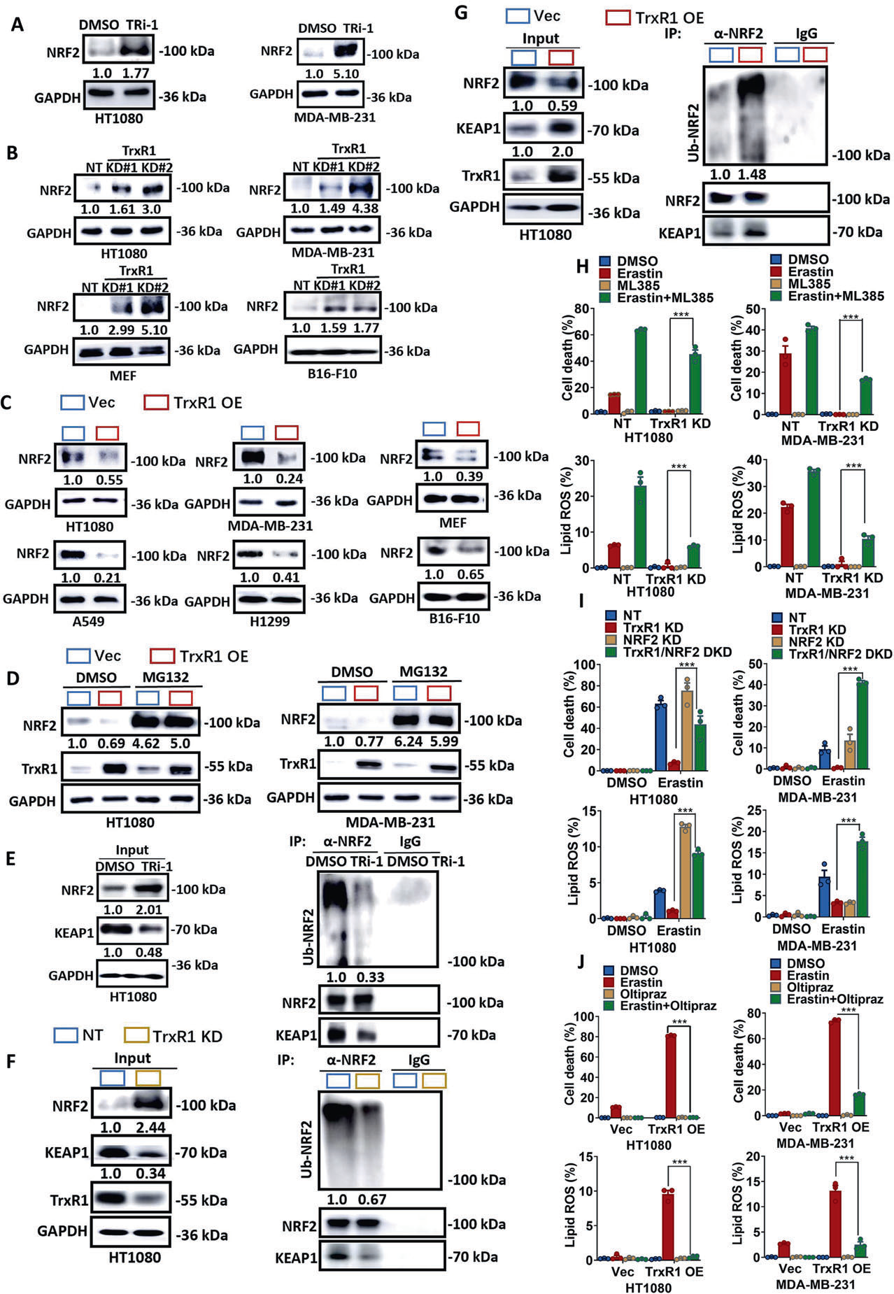
Fig. 3. TrxR1 Promotes Ferroptosis by Decreasing NRF2 Expression. A Treatment with the TrxR1 inhibitor TRi-1 increases the protein level of NRF2 in HT1080 cells and MDA-MB-231 cells. Western blot images and quantitative data confirm the expression of the indicated protein. TRi-1: 2 μM for 12 h. B Knockdown of TrxR1 increases the protein level of NRF2 in multiple cell lines as indicated. Western blot images and quantitative data show the expression of the indicated protein. C Overexpression of TrxR1 suppresses NRF2 expression in multiple cell lines as indicated. Western blot images and quantitative data show the expression of indicated protein. D Treatment with the proteasome inhibitor MG132 blocks downregulation of NRF2 protein induced by TrxR1 overexpression in HT1080 cells and MDA-MB-231 cells. Western blot images and quantitative data show the expression of the indicated protein. MG132: 1 μM for 4 h. E Treatment with the TrxR1 inhibitor TRi-1 suppresses the expression of KEAP1, inhibits the interaction between KEAP1 and NRF2, and decreases the ubiquitination of NRF2. The IP assay was performed using an antibody against NRF2 or an IgG control with the indicated cell lysates. Western blot images and quantitative data show the expression of the indicated protein. F Knockdown of TrxR1 suppresses the expression of KEAP1, inhibits the interaction between KEAP1 and NRF2, and decreases the ubiquitination of NRF2. The IP assay was performed as described in Fig. 3E. Western blot images and quantitative data show the expression of the indicated protein. G Overexpression of TrxR1 increases the expression of KEAP1, enhances the interaction between KEAP1 and NRF2, and promotes the ubiquitination of NRF2. The IP assay was performed as described in Fig. 3E. Western blot images and quantitative data show the expression of the indicated protein. H The NRF2 inhibitor ML385 rescues the ferroptosis sensitivity of TrxR1 KD cells. Cells as indicated were pretreated with 10 μM ML385 for 12 h and then treated with erastin (HT1080: 10 μM; MDA-MB-231: 15 μM) with or without ML385 (10 μM) for 12 h, then cell death and lipid ROS were measured as described in Fig. 1A. I Knockdown of NRF2 rescues the ferroptosis sensitivity of TrxR1 KD cells. Cells as indicated were treated with erastin (HT1080: 10 μM, MDA-MB-231: 15 μM) for 12 h, then cell death and lipid ROS were measured as described in Fig. 1A. J The NRF2 agonist oltipraz suppresses the high ferroptosis sensitivity of TrxR1 overexpressed cells. Cells as indicated were treated with erastin (HT1080: 10 μM, MDA-MB-231: 15 μM) with or without oltipraz (10 μM) for 12 h, then cell death and lipid ROS were measured as described in Fig. 1A. Data are derived from three independent experiments, and each value represents the mean ± SD. P < 0.05, P < 0.01, P < 0.001, t test. (n = 3).(图注取自PDF文本层,来源:Cell Death & Differentiation, 2026)

Fig. 4. TrxR1 promotes ferroptosis by inhibiting NRF2 mediated GPX4 expression. A The NRF2 inhibitor ML385 suppresses the elevated expression level of GPX4 protein in TrxR1 KD cells in HT1080 and MDA-MB-231 cells. Cells as indicated were treated with ML385 (10 μM) for 12 h. Western blot images and quantitative data confirm the expression of the indicated proteins. B The NRF2 inhibitor ML385 suppresses the elevated expression level of GPX4 mRNA in TrxR1 KD cells in HT1080 and MDA-MB-231 cells. Cells as indicated were treated with ML385 (10 μM) for 12 h. RT-qPCR analysis of GPX4 expression is shown. C Knockdown of NRF2 suppresses the elevated expression level of GPX4 protein in TrxR1 KD cells in HT1080 and MDA-MB-231 cells. Western blot images and quantitative data confirm the expression of the indicated proteins. D Knockdown of NRF2 suppresses the elevated expression level of GPX4 mRNA in TrxR1 KD cells in HT1080 and MDA-MB-231 cells. RT-qPCR analysis of GPX4 expression is shown. E The NRF2 agonist oltipraz rescues the expression of GPX4 protein in TrxR1 overexpressed cells in HT1080 and MDA-MB-231 cells. Cells as indicated were treated with oltipraz (10 μM) for 12 h, Western blot images and quantitative data confirm the expression of the indicated proteins. F NRF2 agonist oltipraz rescues the expression level of GPX4 mRNA in TrxR1 KD cells in HT1080 cells and MDA-MB-231 cells. RT-qPCR analysis of GPX4 expression is shown. G A schematic diagram shows NRF2 binding sites on the promoter region of GPX4. H NRF2 binds to the promoter region of GPX4 at two sites, and the TrxR1 inhibitor TRi-1 promotes NRF2 binding on the promoter region of GPX4. The Chromatin-IP assay was performed using an antibody against NRF2 or an IgG control with HepG2 cells treated as indicated. (TRi-1: 2 μM). Data are derived from three independent experiments, and each value represents the mean ± SD. P < 0.05, P < 0.01, P < 0.001, t test. (n = 3).(图注取自PDF文本层,来源:Cell Death & Differentiation, 2026)
第四步:TrxR1 是如何稳定 KEAP1 的?
再往前推一步。KEAP1 的蛋白水平在 TrxR1 敲低后下降,但 mRNA 不变,说明是蛋白稳定性层面的事。蛋白半衰期实验确认了这一点:TrxR1 敲低后 KEAP1 降解变快。MG132 能阻断这个下降,说明是泛素-蛋白酶体途径在起作用。
那到底是哪个 E3 连接酶在给 KEAP1 打泛素标签呢?作者筛选了 8 个 cullin 家族成员(CUL1-9),发现只有 CUL4A 能促进 KEAP1 泛素化。敲低 CUL4A 会稳定 KEAP1、增加其蛋白水平;而过表达 CUL4A 则相反。
他们进一步找到了 KEAP1 上被泛素化的两个赖氨酸位点:K84 和 K312。把这两个位点突变成精氨酸(K84R/K312R),泛素化明显减少,KEAP1 更稳定,NRF2 和 GPX4 下降。有意思的是,双突变更强,说明这两个位点是协同作用的。
@方法论点评:泛素化位点鉴定是功夫活——需要逐个突变候选位点、看泛素化水平变化,同时还要确认突变不影响蛋白本身的折叠和功能(他们用多个突变体相互对照,逻辑严密)。
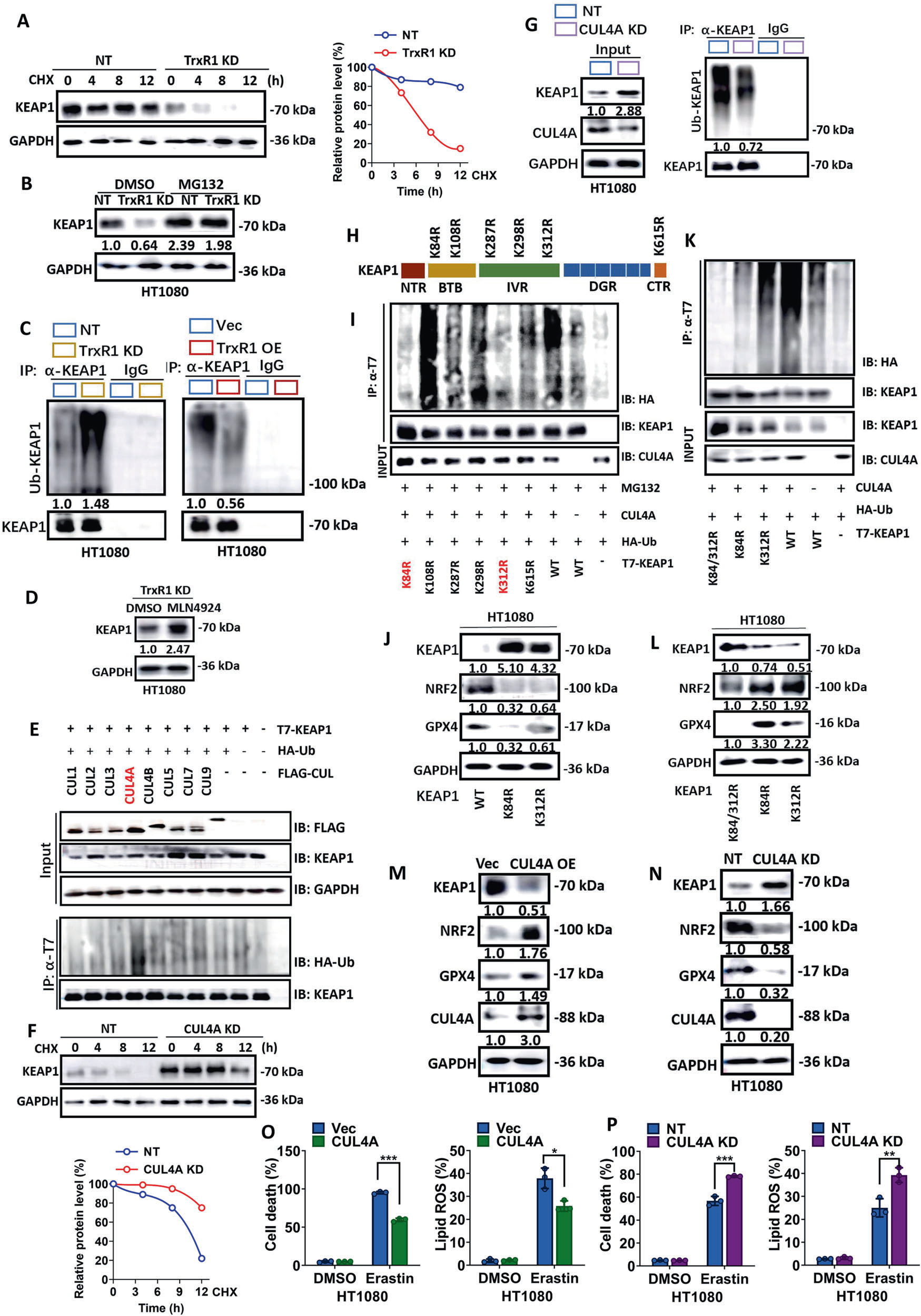
Fig. 5. KEAP1 Is ubiquitinated by CUL4A E3 ligase. A TrxR1 positively regulates the stability of KEAP1. Cells were treated with cycloheximide (CHX, 50 μg/ml) as indicated. Western blot images and quantitative data show the level of the indicated proteins. B Treatment with the proteasome inhibitor MG132 blocks downregulation of KEAP1 protein induced by TrxR1 depletion in HT1080 cells. Western blot images and quantitative data show the expression of the indicated proteins. C TrxR1 negatively regulates the ubiquitination of KEAP1. The IP assay was performed using an antibody against KEAP1 or an IgG control using indicated cell lysates. Ubiquitinated KEAP1 was detected by the antibody against ubiquitin. Western blot images and quantitative data show the expression of the indicated proteins. D Treatment with the protein neddylation inhibitor MLN4924 increases the protein level of KEAP1. HT1080 cells were treated with MLN4924 (10 μM) for 24 h. Western blot images and quantitative data show the expression of the indicated proteins. E CUL4A promotes the ubiquitination of KEAP1. Individual cullin proteins were co-expressed with KEAP1 and ubiquitin in HEK293T cells. The IP assay was performed with an antibody against T7 with indicated cell lysates. Ubiquitinated KEAP1 was detected by the antibody against HA-Ubiquitin. Western blot images show the level of the indicated proteins. F CUL4A negatively regulates the stability of KEAP1. Cells were treated with CHX (50 μg/ml) as indicated. Western blot images and quantitative data show the level of the indicated proteins. G Knockdown of CUL4A suppresses the ubiquitination of KEAP1. The IP assay was performed with an antibody against KEAP1 with indicated cell lysates. Ubiquitinated KEAP1 was detected by the antibody against ubiquitin. Western blot images and quantitative data show the expression of the indicated proteins. H A schematic diagram shows the KEAP1 domains and postion of mutations of lysine. I The K84 and K312 of KEAP1 are ubiquitinated. Individual KEAP1 mutant proteins were co- expressed with CUL4A and ubiquitin in HEK293 cells. Cells were treated with MG132 (0.5 μM) for 24 h. The IP assay was performed using an antibody against T7 with the indicated cell lysates. Ubiquitinated KEAP1 was detected by the antibody against ubiquitin. Western blot images and quantitative data show the expression of the indicated proteins. J The K84R or K312R mutants stabilize KEAP1 and suppress the expression of NRF2 and GPX4. Ubiquitinated KEAP1 was detected by the antibody against ubiquitin. Western blot images and quantitative data show the expression of the indicated proteins. K The K84R and K312R double mutant further decreases the ubiquitination of KEAP1. The IP assay was performed using an antibody against T7 with indicated cell lysates. Ubiquitinated KEAP1 was detected by the antibody against HA-Ubiquitin. L The K84R and K312R double mutant further stabilizes KEAP1 and suppresses the protein level of NRF2 and GPX4. Western blot images and quantitative data show the expression of the indicated proteins. M, N CUL4A negatively regulates the expression of KEAP1 and positively regulates the expression of NRF2 and GPX4. M Knockdown of CUL4A promotes the expression of KEAP1 and suppresses the expression of NRF2 and GPX4 in wildtype HT1080 cells. Western blot images and quantitative data show the expression of the indicated proteins. N Overexpression of CUL4A suppresses the expression of KEAP1 and increases the expression of NRF2 and GPX4 in wildtype HT1080 cells. Western blot images and quantitative data show the expression of the indicated proteins. O Overexpression of CUL4A suppresses the ferroptosis sensitivity in wildtype HT1080 cells. Cells as indicated were treated with erastin (10 μM) for 18 h, then cell death and lipid ROS were measured as described in Fig. 1A. P Knockdown of CUL4A enhances ferroptosis sensitivity in wildtype HT1080 cells. Cells as indicated were treated with erastin (10 μM) for 18 h, then cell death and lipid ROS were measured as described in Fig. 1A. Data are derived from three independent experiments, and each value represents the mean ± SD. P < 0.05, P < 0.01, P < 0.001, t test. (n = 3).(图注取自PDF文本层,来源:Cell Death & Differentiation, 2026)
第五步:CUL4A 的底物适配器是谁?CRBN 浮出水面
CUL4A 需要 DDB1 和底物适配器(DCAF)才能识别底物。沙利度胺是 CRBN 的抑制剂——CRBN 就是 CUL4A 的一个底物适配器。作者发现沙利度胺能阻断 KEAP1 在野生型和 TrxR1 敲低细胞中的降解。敲低 CRBN 同样稳定 KEAP1、减少其泛素化;过表达 CRBN 则相反。
Co-IP 实验确认 CRL4A^CRBN-KEAP1 确实形成一个复合物。结构域 mapping 显示:KEAP1 的 NTR 和 IVR 结构域、CRBN 的 Lon N 端结构域(LNT)是它们互作的关键区域。
功能上,敲低 CRBN 让细胞对 erastin 更敏感,沙利度胺也有同样效果。所以,CRBN 是负责 KEAP1 泛素化的 E3 连接酶的底物识别亚基。

Fig. 6. CRBN Is the substrate adaptor protein for CUL4A-mediated KEAP1 Ubiquitination. A Treatment with the neddylation inhibitor MLN4924 or the CRBN inhibitor Thalidomine increases the protein level of KEAP1. HT1080 cells were treated with MLN4924 (10 μM) for 24 h or thalidomine (10 μM) for 24 h. Western blot images and quantitative data show the expression of the indicated proteins. B CRBN negatively regulates the stability of KEAP1. Cells were treated with CHX (50 μg/ml) as indicated. Western blot images and quantitative data show the level of the indicated proteins. C Knockdown of CRBN suppresses the ubiquitination of KEAP1 and the interaction between KEAP1 and CUL4A. The IP assay was performed using an antibody against KEAP1 with the indicated cell lysates. Ubiquitinated KEAP1 was detected by the antibody against ubiquitin. Western blot images and quantitative data show the expression of the indicated proteins. D Overexpression of CRBN promotes the ubiquitination of KEAP1 and the interaction between KEAP1 and CUL4A. The IP assay was performed using an antibody against KEAP1 with the indicated cell lysates. Ubiquitinated KEAP1 was detected by the antibody against ubiquitin. Western blot images and quantitative data show the expression of the indicated proteins. E The NTR and IVR domains of KEAP1 are required for the interaction between KEAP1 and CRBN. F The Lon N-terminal (LNT) domain of CRBN is required for the interaction between KEAP1 and CRBN. G Knockdown of CRBN promotes the expression of KEAP1 and suppresses the expression of NRF2 and GPX4 in wildtype HT1080 cells. Western blot images and quantitative data show the expression of the indicated proteins. H Thalidomide treatment sensitizes erastin induced ferroptosis. B16-F10 or HT1080 cells were treated as indicated for 16 h, then cell death and lipid ROS were measured as described in Fig. 1A. erastin: 10 μM, Thalidomide: 10 μM, Fer-1: 2 μM. I Knockdown of CRBN enhances ferroptosis sensitivity in wildtype HT1080 cells. Cells as indicated were treated with erastin (10 μM) for 18 h, then cell death and lipid ROS were measured as decribed in Fig. 1A. Data are derived from three independent experiments, and each value represents the mean ± SD. P < 0.05, P < 0.01, P < 0.001, t test. (n = 3).(图注取自PDF文本层,来源:Cell Death & Differentiation, 2026)
第六步:TrxR1 如何"踩住"CRL4A^CRBN 这个刹车?
TrxR1 敲低时,CUL4A 的蛋白水平上升,同时 KEAP1 与 CUL4A-CRBN 的互作增强;过表达 TrxR1 则相反。而且 TrxR1 还影响 CRL4A^CRBN 的其他底物(如 IKZF3 和 CK1α)——说明 TrxR1 不是只针对 KEAP1,而是对整个复合物的活性有广泛影响。
机制上,作者发现 TrxR1 直接与 CRBN 的 LNT 结构域结合,而 KEAP1 也是通过同一个结构域与 CRBN 互作的。这就构成了一个竞争关系:TrxR1 过表达时,它和 KEAP1 “抢” CRBN,导致 KEAP1 被 CRBN 识别减少、泛素化降低、稳定性增加。反过来,TrxR1 缺失时,KEAP1 更容易被 CRBN 抓住并降解。
但注意,TrxR1 的 U498 硒代半胱氨酸突变体虽然还能结合 CRBN,却无法稳定 KEAP1 和降低 NRF2/GPX4——说明 TrxR1 对 KEAP1 的调控至少有两层机制:一是通过竞争性结合 CRBN,二是 U498 还参与了其他未知的调控环节(可能涉及 TrxR1 的氧化还原活性)。
@方法论点评:竞争性结合这个概念非常关键。他们用"同一个互作界面"来解释 TrxR1 如何不直接泛素化 KEAP1 却能稳定它——这种"间接调控"的思路在信号通路研究中很常见,但需要有直接的 Co-IP 和 domain mapping 数据支撑。

Fig. 7. TrxR1 Stabilizes KEAP1 and Promotes Ferroptosis by Suppressing CRL4ACRBN E3 Complex. A, B TrxR1 negatively regulates the expression of CUL4A and the formation of CUL4A-CRBN-KEAP1 complex. A Knockdown of TrxR1 promotes the expression of CUL4A and the formation of CUL4A -CRBN-KEAP1 complex. The IP assay was performed using an antibody against KEAP1 or CRBN with the indicated cell lysates. Western blot images and quantitative data show the expression of the indicated proteins. B Overexpression of TrxR1 suppresses the expression of CUL4A and the formation of CUL4A-CRBN-KEAP1 complex. The IP assay was performed using an antibody against KEAP1 or CRBN with indicated cell lysates. Western blot images and quantitative data show the expression of the indicated proteins. C TrxR1 broadly modulates the stability of CRL4ACRBN substrates. Western blot images and quantitative data show the expression of the indicated proteins. D Knockdown of CUL4A restores the expression of KEAP1 and suppresses the upregulation of the expression of NRF2 and GPX4 in TrxR1 KD HT1080 cells. Western blot images and quantitative data show the expression of the indicated proteins. E Overexpression of CUL4A suppresses the upregulation of KEAP1 expression and restores the downregulation of the expression of NRF2 and GPX4 in TrxR1 OE HT1080 cells. Western blot images and quantitative data show the expression of the indicated proteins. F Knockdown of CUL4A restores the ferroptosis sensitivity of TrxR1 KD cells. Cells as indicated were treated with erastin (10 μM) for 18 h, then cell death and lipid ROS were measured as described in Fig. 1A. G Overexpression of CUL4A suppreses the higher ferroptosis sensitivity in TrxR1 OE cells. Cells as indicated were treated with erastin (10 μM) for 18 h, then cell death and lipid ROS were measured as described in Fig. 1A. H Inhibition of CRBN by thalidomide restores the protein level of KEAP1 and suppresses the upregulation of NRF2 and GPX4 in TrxR1 KD HT1080 cells. Indicated cells were treated with thalidomide (10 μM) for 24 h. Western blot images show and quantitative data the expression of the indicated proteins. I Knockdown of CRBN restores the protein level of KEAP1 and suppresses the upregulation of NRF2 and GPX4 in TrxR1 KD HT1080 cells. Western blot images show and quantitative data the expression of the indicated proteins. J Knockdown of CRBN restores the ferroptosis sensitivity of TrxR1 KD cells. Cells as indicated were treated with erastin (10 μM) for 18 h, then cell death and lipid ROS were measured as decribed in Fig. 1A. K, L TrxR1 is not a substrate of CRL4ACRBN E3 complex. Western blot images show the expression of the indicated proteins. K Overexpression of CUL4A does not change the protein level of TrxR1. Western blot images show the expression of the indicated proteins. L Overexpression of CRBN does not change the protein level of TrxR1. Western blot images show the expression of the indicated proteins. M TrxR1 interacts specifically with CRBN but not with other components of CUL4A-CRBN-KEAP1 complex. The IP assay was performed using specific antibody against TrxR1 as described in Fig. 3E. Western blot images show the expression of the indicated proteins. N TrxR1 interacts with the Lon N-terminal (LNT) domain of CRBN. The IP assay was performed using specific antibody against Myc tag as described in Fig. 3E. Western blot images show and quantitative data the expression of the indicated proteins. O More than one domain of TrxR1 is required for its interaction with CRBN. The IP assay was performed using specific antibody against HA tag as described in Fig. 3E. Western blot images show and quantitative data the expression of the indicated proteins. P TrxR1 promotes KEAP1 stabilization by competitively binding to CRBN. Western blot images show and quantitative data the expression of the indicated proteins. Q The selenocysteine U498S mutant of TrxR1 does not impair its interaction with CRBN. The IP assay was performed using specific antibody against HA tag as described in Fig. 3E. Western blot images show and quantitative data the expression of the indicated proteins. Data are derived from three independent experiments, and each value represents the mean ± SD. P < 0.05, P < 0.01, P < 0.001, t test. (n = 3).(图注取自PDF文本层,来源:Cell Death & Differentiation, 2026)
第七步:体内验证和临床转化可能性
最后,作者把这一整套机制搬到小鼠身上。B16-F10 黑色素瘤异种移植模型中,高表达 TrxR1 的肿瘤对铁死亡诱导剂 IKE(erastin 的体内可用版本)更敏感,肿瘤生长抑制更明显,同时 KEAP1 升高、NRF2 和 GPX4 降低、脂质过氧化标志物 4-HNE 升高。反过来,TrxR1 敲低的肿瘤对 IKE 耐药。
更值得关注的是,沙利度胺(CRBN 抑制剂,FDA 已批准用于多发性骨髓瘤)联合 IKE 治疗,能显著抑制肿瘤进展,比单药效果好。IHC 验证了联合组 KEAP1 升高、NRF2/GPX4 降低、4-HNE 升高。
TCGA 数据还显示,TrxR1 在多种肿瘤中高表达,且高表达与患者总生存期差相关——这为 TrxR1 作为铁死亡诱导疗法的生物标志物提供了临床层面的依据。
@方法论点评:体内实验是这篇文章从"分子机制"到"临床前转化"的关键一跃。用 IKE(erastin 的改良版)是因为普通 erastin 在体内代谢太快、药代动力学差;用 4-HNE 作为铁死亡的 IHC 标志物也是当前领域的常规做法。
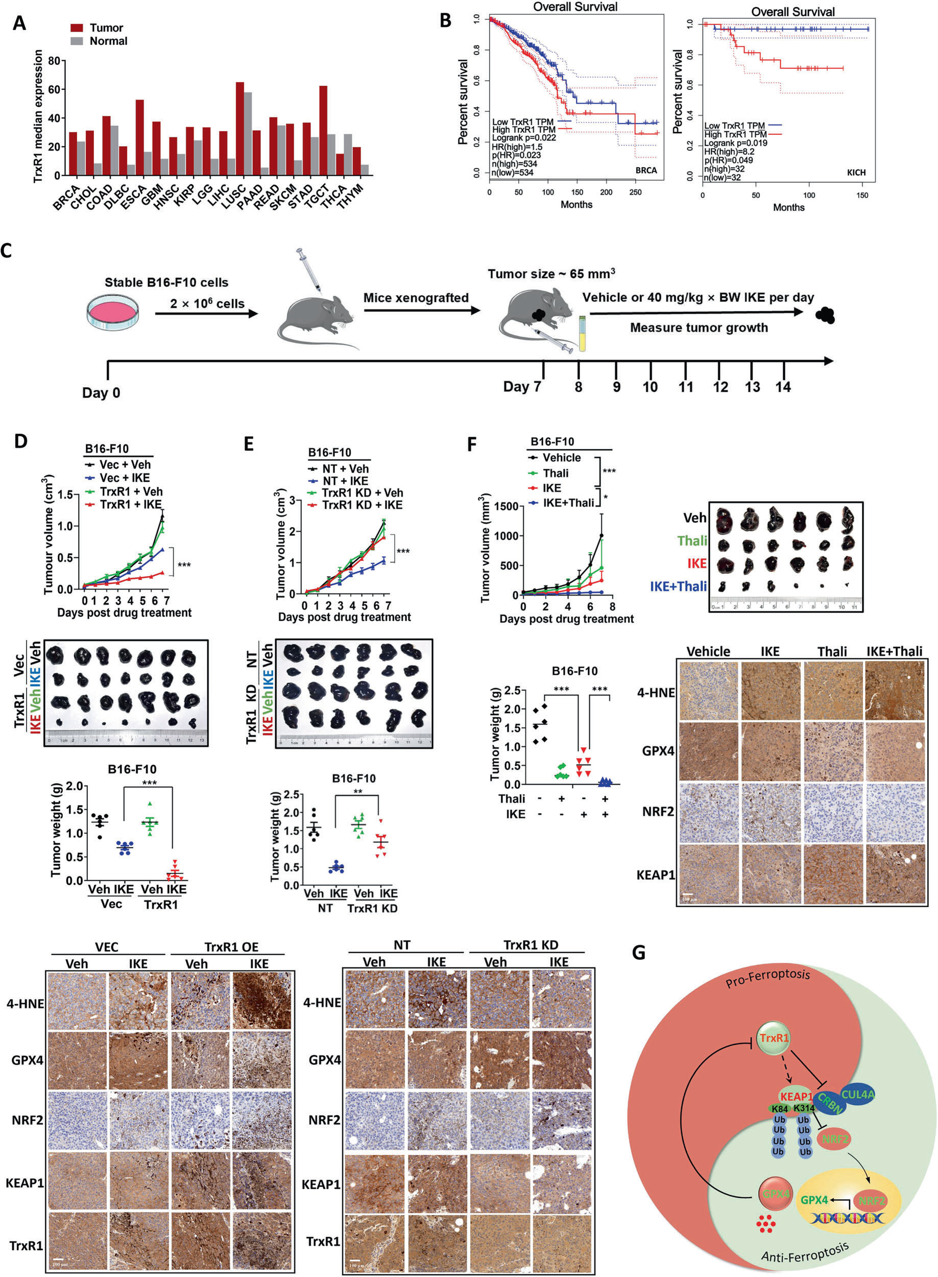
Fig. 8. High expression of TrxR1 sensitizes cancer cell to ferroptosis in vivo. A Tumor tissues exhibit elevated expression of TrxR1 compared to adjacent normal tissues in multiple cancer patients. BRCA (Breast Invasive Carcinoma), CHOL (Cholangiocarcinoma), COAD (Colorectal Adenocarcinoma), DLBC (Diffuse Large B-cell Lymphoma), ESCA (Esophageal Carcinoma), GBM (Glioblastoma Multiforme), HNSC (Head and Neck Squamous Cell Carcinoma), KIRP (Kidney Renal Papillary Cell Carcinoma), LGG (Low Grade Glioma), LIHC (Liver Hepatocellular Carcinoma), LUSC (Lung Squamous Cell Carcinoma), PAAD (Pancreatic Adenocarcinoma), READ (Rectal Adenocarcinoma), SKCM (Skin Cutaneous Melanoma), STAD (Stomach Adenocarcinoma), TGCT (Testicular Germ Cell Tumor), THCA (Thyroid Cancer), THYM (Thymoma). B Higher expression of TrxR1 is correlated with poor overall survival in multiple different types of cancer patients. BRCA (Breast Cancer), KICH (Kidney Chromophobe). C A schematic diagram shows the procedure of the mouse xenograft model experiment. D Overexpression of TrxR1 sensitizes cancer cells to ferroptosis-inducing tumor therapy in a mouse xenograft model. Mouse xenograft model experiments with indicated stable B16-F10 cells were performed as described in Fig. 8C. Tumor volume was measured daily. Tumor volume post-IKE treatment is shown and tumors were photographed and weighted after 7 days IKE treatment. The expression of indicated protein or 4-HNE in tumor tissue were analyzed by IHC assay using specific antibodies. Scale bar: 100 μm. E Knockdown of TrxR1 confers resistance to ferroptosis-inducing tumor therapy in a mouse xenograft model. Mouse xenograft model experiments with indicated stable B16-F10 cells were performed as described in Fig. 8C and tumor measured as described in Fig. 8D. The expression of indicated protein or 4-HNE in tumor tissue were analyzed by IHC assay using specific antibodies. Scale bar: 100 μm. F Thalidomide and IKE combination treatment results in markedly retarded tumor progession. Mouse xenograft model experiments with B16-F10 cells were performed as described in Fig. 8c and tumor measured as described in Fig. 8d. Thalidomide: 40 mg/kg, IKE: 40 mg/kg. Scale bar: 100 μm. G The working model showing how TrxR1 promotes ferroptosis by inhibiting CRL4ACRBN E3 ligase complex mediated KEAP1 ubiquitination and degradation, thereby suppressing NRF2-dependent GPX4 expression. The average values are present in the bar graphs (means ± SD) (n ≥6 for each group). P < 0.05, P < 0.01, P < 0.001, t test. (n ≥6).(图注取自PDF文本层,来源:Cell Death & Differentiation, 2026)
核心结论
作者最终提出的模型是:TrxR1 通过两方面的作用稳定 KEAP1——一方面抑制 CUL4A 表达,另一方面与 KEAP1 竞争结合 CRBN(CUL4A 的底物适配器)。KEAP1 稳定后,NRF2 被更多泛素化降解,导致 GPX4 转录下降,细胞对铁死亡的敏感性增加。
这个发现之所以重要,是因为它打破了"抗氧化系统=抑制铁死亡"的简单认知。TrxR1 虽然是抗氧化酶,但在铁死亡这个特定场景下,它通过调控 GPX4 的表达而成为铁死亡的"加速器"。文章还用"阴阳"来比喻 TrxR1 和 GPX4 的互作关系——GPX4 抑制 TrxR1 二聚化,TrxR1 抑制 GPX4 表达,形成一种动态平衡。
对耐药/DTP/PGCC 的启示
TrxR1 作为铁死亡敏感性的生物标志物:既然 TrxR1 高表达让癌细胞对铁死亡诱导剂敏感,那么在 TrxR1 高表达的耐药肿瘤中,联合铁死亡诱导剂可能是一种策略。尤其是在那些对常规化疗耐药的 DTP(药物耐受持久细胞)中,如果 TrxR1 表达水平高,或许可以"趁其虚弱"用铁死亡清除。 沙利度胺的"老药新用":沙利度胺本身是 CRBN 抑制剂,已用于多发性骨髓瘤。这篇文章为沙利度胺联合铁死亡诱导剂提供了机制上的"合理性论证"。对那些 CRBN 表达尚存的肿瘤,沙利度胺可以增强铁死亡敏感性,这为临床组合方案的设计提供了新思路。 对多倍体巨细胞(PGCC)的可能影响:PGCC 通常对多种治疗方式耐受,其抗氧化能力往往增强。如果 PGCC 中 TrxR1 表达下调或功能受损(文中未讨论,这是一个推测空间),可能导致 GPX4 上调、铁死亡耐受,需要进一步验证。反过来,如果 TrxR1 高表达,PGCC 可能反而对铁死亡敏感。这个问题文中未涉及,但值得后续探索。
局限
机制研究主要在 HT1080、MDA-MB-231 等少数细胞系中进行,是否在所有癌种中通用有待扩展。 NRF2 调控的下游基因远不止 GPX4(还包括 GCLC、GCLM、FTH1 等),作者把效应归因于 GPX4 下调,但其他 NRF2 靶基因的贡献没有系统排除。 TrxR1 如何负调控 CUL4A 的蛋白水平,文中没有深究。 TrxR1 如何负调控 NRF2 的 mRNA 水平,机制也未阐明。 体内实验用的是免疫缺陷小鼠或同种移植模型,尚未在更接近临床的 PDX 模型或免疫健全模型中验证。
来源
期刊:Cell Death & Differentiation,2026。DOI: 10.1038/s41418-026-01691-z