一句话亮点
这篇文章发现,PHB2通过结合并激活脂代谢酶ACSL3,促进单不饱和脂肪酸(MUFA)掺入膜磷脂,从而抑制铁死亡、驱动胃癌顺铂耐药;而FDA已批准的CXCR4拮抗剂Mavorixafor能打断这个蛋白-蛋白相互作用,将"耐药"的膜脂组成"拨回"易铁死亡状态,在体内模型里重新增敏顺铂。
背景/痛点
铂类是胃癌化疗的基石,但耐药几乎是宿命。经典机制——DNA修复增强、药物外排、凋亡逃逸——已经研究了很多年,但不足以解释临床耐药的顽固性。所以作者把视线投向非凋亡性细胞死亡,尤其铁死亡。铁死亡的关键在于膜脂上多不饱和脂肪酸(PUFA)被氧化,而单不饱和脂肪酸(MUFA)的掺入能"踩刹车",减少脂质过氧化底物。ACSL4是PUFA激活酶,促铁死亡;ACSL3是MUFA激活酶,抑铁死亡。ACSL4上游研究不少,但ACSL3怎么被调控、在耐药里扮演什么角色,并不清楚。PHB2以前主要被认为在线粒体里干杂活(维持结构、参与线粒体自噬),有没有胞质里的"兼职功能"?这就是本文切入的窗口。
推理链分步拆解
Step 1:先找耐药里冒头的分子——PHB2是怎么被"捞"出来的
作者先做了一个很经典的耐药模型构建:体外筛选顺铂耐药的AGS细胞,再植回小鼠肚子里形成CDDP-R xenograft,然后RNA-seq对比敏感和耐药肿瘤的转录组。差异基因一堆,怎么聚焦?他们把自家差异基因和NCI-60 CellMiner里基因表达与顺铂GI50的相关性做交集,从中"捞"出PHB2——mRNA在耐药瘤里高表达,而且在NCI-60里表达越高、顺铂越不敏感。紧接着在病人组织里用IHC验证,蛋白水平也是耐药组更高。到这里问题来了:是伴随现象,还是真的驱动耐药?他们用shRNA敲低和过表达做双向因果验证,敲低后耐药细胞重新变敏感,过表达后敏感细胞变耐药;体内也一样,敲低PHB2后顺铂压不住瘤了。`@方法论点评:这里值得学的是"双筛策略"——自己的组学差异 + 公共数据库的药物敏感性关联,两者交集能显著缩小候选范围,避免只看差异基因的假阳性。再看双向表型(敲低+过表达)确认因果关系,是功能研究的金标准。"
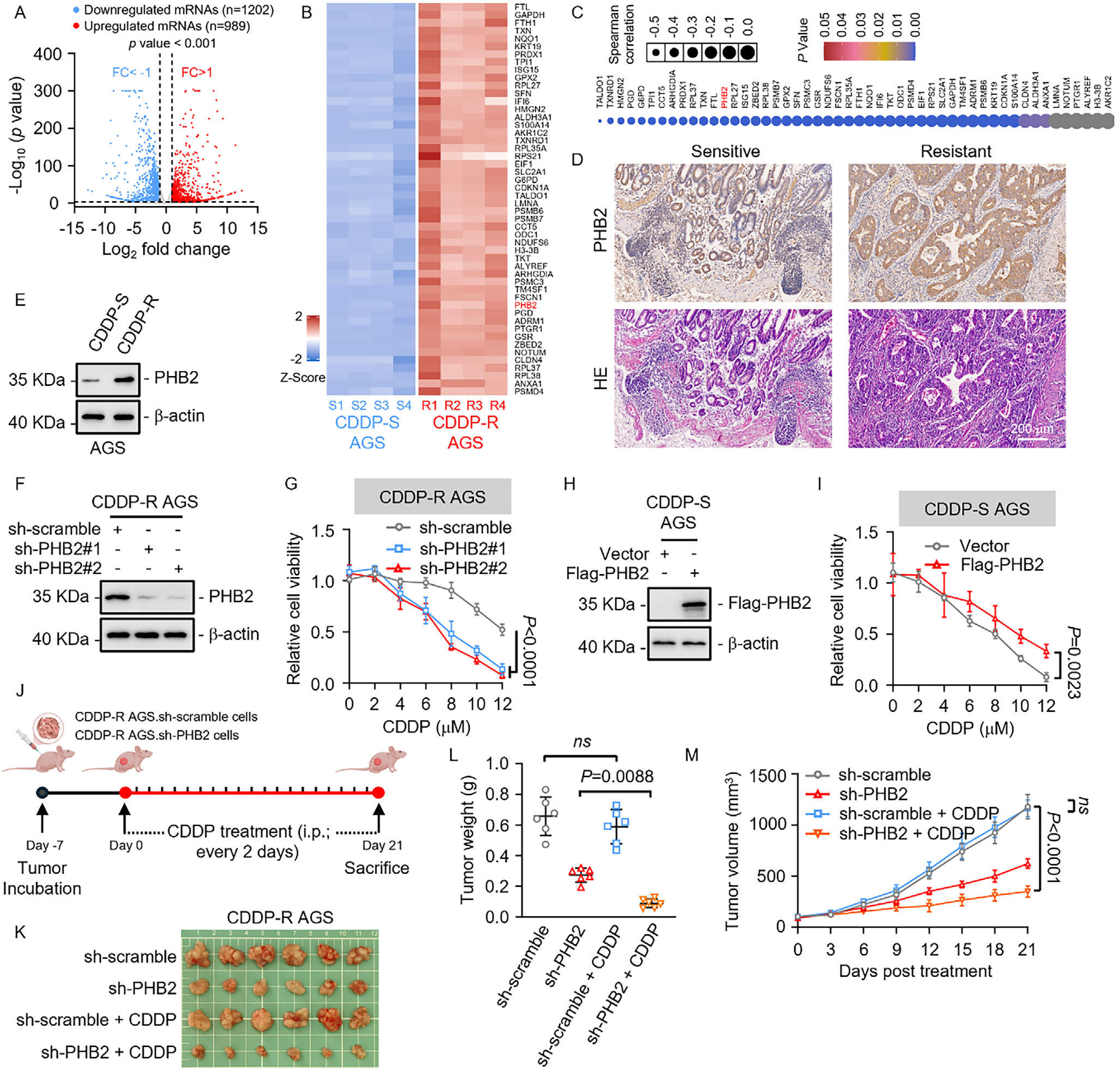
Fig. 1. High PHB2 expression promotes chemotherapy resistance in GC. A,B) Volcano plot (A) of differentially expressed proteins and heatmap (B) of the top 50 proteins in CDDP-R versus CDDP-S AGS xenograft tumors (fold change > 1 or < –1, P < 0.001; n = 4 mice per group). C) Dot plot showing the correlation between expression of selected genes and CDDP sensitivity (GI50 values) across the NCI-60 cell line panel, based on CellMiner analysis. Each dot represents a gene, with size indicating the Spearman correlation coefficient and color corresponding to the statistical significance ( P value). Genes are ranked by correlation strength. PHB2 is highlighted in red, exhibiting a strong negative correlation with CDDP sensitivity. D) IHC staining analysis of PHB2 expression in GC tissues from cisplatin-sensitive (CDDP-S, n = 10) and cisplatin-resistant (CDDP-R, n = 24) patient groups. Representative images are shown. Scale bar, 200 µm. E) Western blot analysis of PHB2 expression in CDDP-S and CDDP-R AGS cells. β-actin was used as control. F,G) Western blot examined PHB2 knockdown efficiency in CDDP-R AGS cells transfected with sh-PHB2 (F), and subsequent MTT assay showed reduced cell viability after 72 hours of CDDP treatment (G). H,I) Overexpression of PHB2 (H) enhanced resistance to CDDP-induced cytotoxicity, as assessed by MTT assay after 72 hours of CDDP treatment (I). J–M) Schematic illustration of the establishment of CDDP-R AGS.sh-scramble and AGS.sh-PHB2 xenograft tumors. BALB/c nude mice bearing CDDP-R AGS-derived xenografts were treated with either 0.9% saline or CDDP (5 mg/kg, intraperitoneally [i.p.]) every 2 days for 3 weeks. n = 6 mice per group (J). Representative images (K), tumor weight (L), and growth curves (M) of AGS.sh-scramble and AGS.sh-PHB2 xenografts in BALB/c nude mice. Scale bar, 1 cm. Data represent three independent experiments and are presented as mean ± SD. Quantitative data were normalized to the corresponding sh-scramble control (G,I). One-way (L) or two-way ANOVA followed by Tukey’s multiple comparison (G, I, M).(图注取自PDF文本层,来源:Advanced Science, 2026)
Step 2:铁死亡通路冒出来了——是"主犯"还是"围观群众"?
KEGG富集分析显示,耐药肿瘤里铁死亡通路被显著抑制。但他们没停在富集分析层面,而是做了一步很关键的"排他性验证":用一系列细胞死亡通路抑制剂去"救"PHB2敲低造成的细胞死亡。结果只有铁死亡抑制剂(Ferrostatin-1和Liproxstatin-1)能救,凋亡、坏死、自噬的抑制剂都无效。这就把PHB2和铁死亡的特异性关联坐实了。接下来他们测了脂质过氧化的系列指标——Liperfluo、C11-BODIPY、MDA、电镜——全部指向PHB2敲低后铁死亡增强。`@方法论点评:这一步的"抑制剂救援实验"非常关键。如果你只看到PHB2敲低后铁死亡相关基因变化,那可能只是相关性;但用不同死亡通路的抑制剂去"反推"死亡方式,能直接锁定铁死亡是这个表型的主要执行者,而不是伴随现象。"
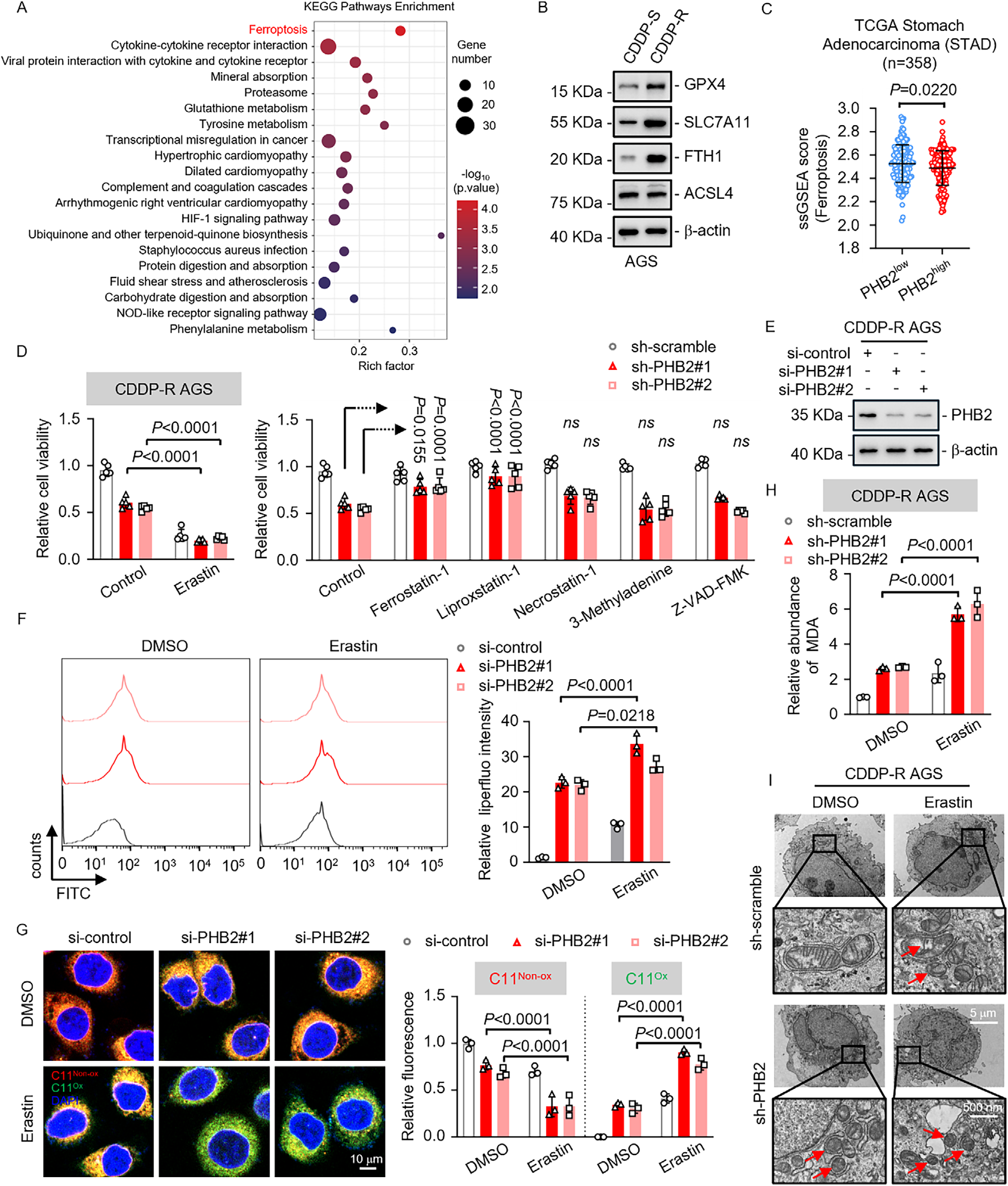
Fig. 2. PHB2 drives chemotherapy resistance in GC by regulating ferroptosis. A) KEGG pathway enrichment analysis revealed that ferroptosis was significantly enriched in CDDP-S compared to CDDP-R AGS xenograft tumors. Dot size represents the number of genes in each KEGG pathway; KEGG, Kyoto Encyclopedia of Genes and Genomes. B) Western blot analysis of ferroptosis-related genes GPX4, SLC7A11, FTH1, and ACSL4 in CDDP-S and CDDP-R AGS cells, with β-actin used as control. C) ssGSEA scores for the ferroptosis pathway in low and high PHB2 expression clusters in TCGA Stomach Adenocarcinoma (STAD) cohort (n = 358). D) CDDP-R AGS cells were treated for 72 hours with pathway-specific compounds, including the ferroptosis inducer Erastin; ferroptosis inhibitors Ferrostatin-1 and Liproxstatin-1; the necroptosis inhibitor Necrostatin-1; the autophagy inhibitor 3- Methyladenine; and the apoptosis inhibitor Z-VAD-FMK. Cell viability was then evaluated by MTT assay. E–G) Western blot analysis confirmed PHB2 knockdown efficiency in CDDP-R AGS cells transfected with si-PHB2 (E). Lipid peroxidation, assessed using Liperfluo (F) and BODIPY 581/591 C11 (G) staining, was elevated following PHB2 knockdown, especially in the Erastin-treated group. H) MDA level analysis revealed a significant increase(图注取自PDF文本层,来源:Advanced Science, 2026)
Step 3:怎么找到搭档ACSL3?——免疫沉淀+质谱"钓鱼"
既然PHB2调控脂质过氧化,但本身不是代谢酶,它很可能通过蛋白-蛋白相互作用"借力"来干活。他们用IP-MS钓PHB2互作蛋白,结果里有一堆脂代谢酶,其中ACSL3最显眼——而且ACSL3刚好是MUFA激活酶,和前面铁死亡抑制的方向吻合。Co-IP和免疫荧光都确认了二者在胞质共定位。接着用截短体做"分区拆解",发现PHB2的C端负责结合ACSL3,而ACSL3的AMP-binding domain是结合PHB2的关键区域。更有意思的是,他们用结构预测找到了三个关键残基(W244/H254/E260),突变掉任何一个都会削弱结合、同时增加脂质过氧化。`@方法论点评:这里有一个容易被忽视的点——他们不是直接看ACSL3表达是否受PHB2调控(事实是表达不变),而是直接测ACSL3活性(MUFA含量、OA-alkyne incorporation)。这提醒我们:蛋白互作可以改变酶活性而不是表达水平,如果只做WB可能就漏掉机制了。"

Fig. 3. PHB2 suppresses ferroptosis by binding to the AMP-binding domain of ACSL3. A) PHB2-interacting enzymes identified by IP- MS analysis, highlighting those involved in lipid metabolism and oxidative stress pathways in the table. B) Endogenous PHB2 and ACSL3 co-immunoprecipitated in AGS and MKN-28 cells. C) Co-IP of exogenous Flag-PHB2 and HA-ACSL3 in HEK-293T cells. D) Representative immunofluorescence images showing co-localization of PHB2 and ACSL3 in AGS cells. Scale bar, 10 µm. E,F) Deletion-mapping experiments revealed(图注取自PDF文本层,来源:Advanced Science, 2026)
Step 4:PHB2-ACSL3如何重塑膜脂?——MUFA/PUFA比例的"拨盘"
敲低PHB2后ACSL3蛋白量没变,但MUFA(油酸)水平下降,PUFA(亚油酸)不变,说明ACSL3的活性被PHB2"增强"了。他们用OA-alkyne + click chemistry直接"看"MUFA被掺入膜的情况,PHB2敲低后荧光明显减弱。更关键的是脂质组学:PHB2敲低后,含MUFA的PC和PE显著下降,但含PUFA的PC/PE不受影响。这就形成了一条清晰的逻辑链:PHB2结合ACSL3的AMP-binding domain → 增强ACSL3活性 → 更多MUFA被活化成MUFA-CoA → 掺入膜磷脂 → MUFA/PUFA比例升高 → 膜不易被氧化 → 铁死亡耐受。反向验证也很漂亮:敲低ACSL3后,再强行过表达PHB2也救不回MUFA掺入和铁死亡抑制,说明PHB2必须通过ACSL3才能起作用。`@方法论点评:这篇的机制验证层层递进,从"谁和谁结合"到"结合在哪个结构域"到"结合后改变什么酶活性"到"酶活性改变后影响什么脂质类别",每一步都有截短体/点突变/敲除回复等正交方法锁定,而不是只靠单一手段下结论。"
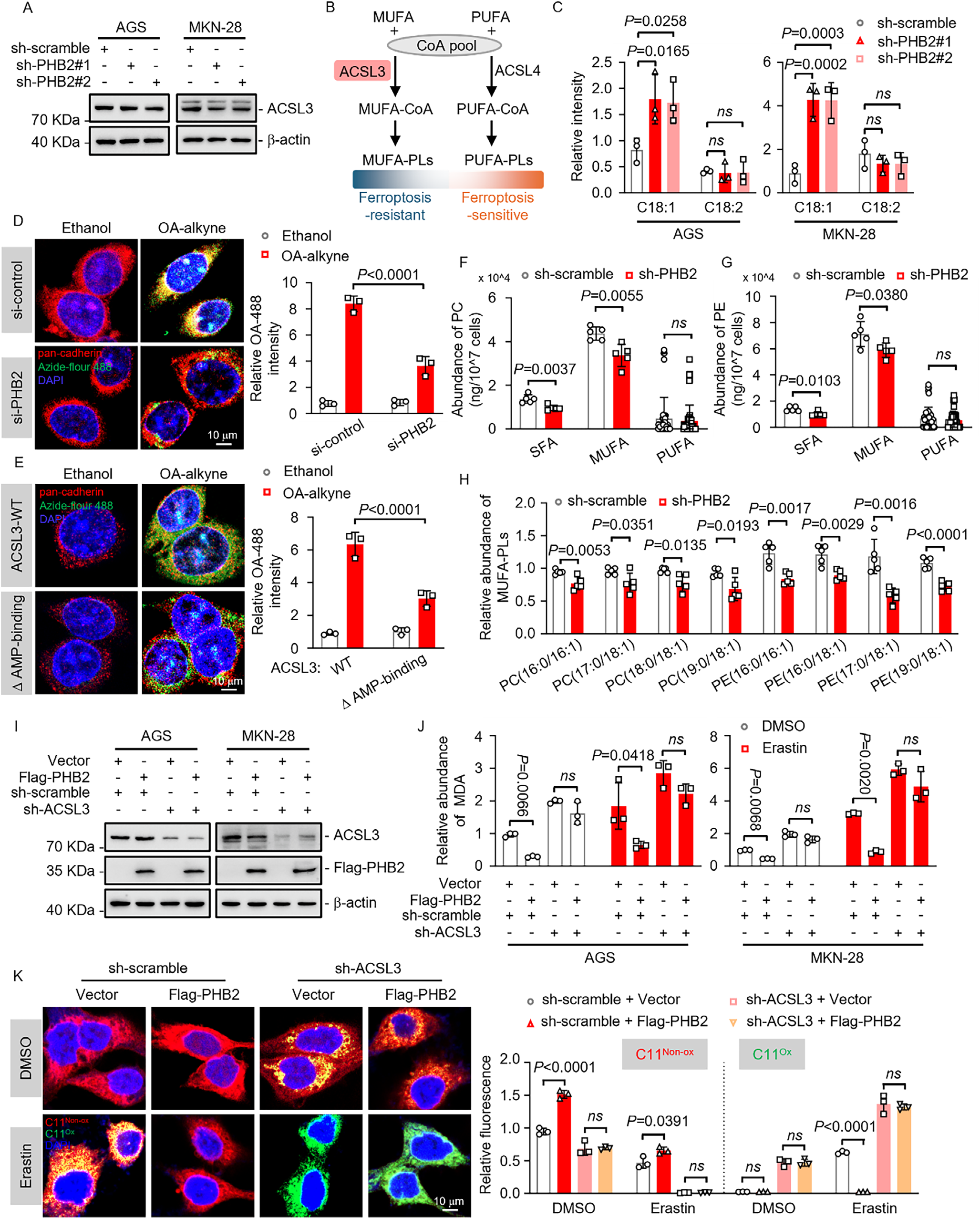
Fig. 4. PHB2 regulates ACSL3 activities controlling the MUFA-to-PUFA ratio. A) Western blot analysis showing ACSL3 expression levels in AGS.sh-PHB2 and MKN-28.sh-PHB2 cells. B) Schematic illustrating ACSL3-mediated regulation of membrane phospholipid remodeling during ferroptosis. C) Quantification of cellular MUFA (OA; C18:1) and PUFA (LA; C18:2) levels by targeted lipidomics using LC-MS/MS in AGS.sh-PHB2 and MKN-28.sh-PHB2 cells (n = 3 independent samples). D) IF staining and quantification of membrane-associated OA-488 fluorescence intensity in AGS.si-control and AGS.si-PHB2 cells treated with either ethanol (vehicle control) or OA-alkyne. Cells were treated with 20 µM OA-alkyne for 10 hours, followed by a 10-hour chase in regular medium. pan-cadherin used as a plasma membrane marker. Scale bar, 10 µm. E) OA-488 fluorescence intensity(图注取自PDF文本层,来源:Advanced Science, 2026)
Step 5:老药新用的"神操作"——虚拟筛选+SPR锁定Mavorixafor
既然PHB2-ACSL3互作是驱动耐药的关键节点,能不能用药物去打断它?他们做了基于结构的虚拟筛选,从7000多个FDA已上市药物(TargetMol T001库)里找能结合ACSL3 AMP-binding domain的小分子,经过MMGBSA、PLIF、结构多样性筛选,再经过SPR亲和力验证,最后锁定了Mavorixafor——一个已经被批用于WHIM综合征的CXCR4拮抗剂。紧接着一系列功能验证:Mavorixafor能打断内源性PHB2-ACSL3互作(Co-IP确认)、能逆转MUFA掺入、能增加PUFA磷脂、能诱导脂质过氧化、能被铁死亡抑制剂逆转。体内CDX和PDO模型里,Mavorixafor + 顺铂的组合显著抑制耐药肿瘤生长,而加上Liproxstatin-1后效果消失,确认了铁死亡依赖性。`@方法论点评:这是典型的"老药新用"策略,但亮点在于它不是随便碰运气筛选细胞毒性,而是基于清晰的结构生物学假设去"定向筛选",再用SPR验证物理结合,最后用遗传学手段(敲低PHB2或ACSL3)确认靶点特异性。这种"从机制到药物"的正向转化路径,比"筛到药再找靶点"更具说服力。"

Fig. 5. Mavorixafor targets the PHB2-ACSL3 interaction to regulate ACSL3-dependent phospholipid remodeling and overcome ferroptosis resistance in GC. A) Virtual screening of the TargetMol-T001 library, ranked by MMGBSA binding free energy, identified Mavorixafor as a potential inhibitor of the PHB2-ACSL3 interaction. The chemical structure of Mavorixafor is shown on the right. B) Surface plasmon resonance (SPR) analysis of the binding affinity between immobilized recombinant human ACSL3 and serially diluted Mavorixafor (KD = 6.374 ×10− 6 M). C) Binding conformation(图注取自PDF文本层,来源:Advanced Science, 2026)
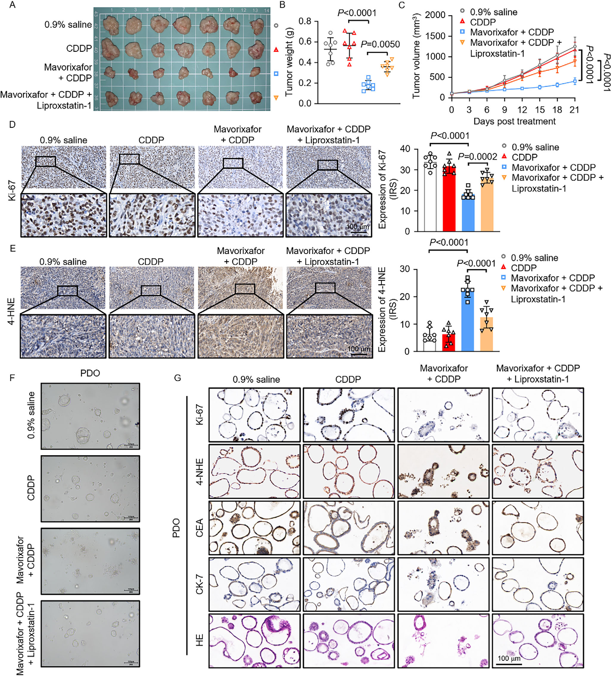
Fig. 6. Mavorixafor sensitizes drug-resistant GC via ferroptosis in CDX and PDO models. A–C) Representative image (A), tumor weight (B), and growth curves (C) of CDDP-R AGS xenografts treated with 0.9% saline (control), CDDP, Mavorixafor combined with CDDP, and Mavorixafor combined with CDDP and Liproxstatin-1 in BALB/c nude mice. Scale bar, 1 cm. D, E) IHC staining and quantification of Ki-67 (D) and 4-HNE (E) in CDDP-R AGS xenograft tumors with the indicated treatments. Representative IHC images are shown. Scale bar, 100 µm. IRS: Immunoreactive score. F) Representative bright-field images of CDDP-R GC PDOs treated with 0.9% saline (control), CDDP, Mavorixafor combined with CDDP, and Mavorixafor combined with CDDP and Liproxstatin-1. G) Representative IHC staining of Ki-67, 4-HNE, CEA, and CK-7, as well as HE staining, in CDDP-R GC PDOs under the indicated treatments. Scale bar, 100 µm. Data represent three independent experiments and are presented as mean ± SD. One-way (B,D,E) or two-way ANOVA followed by Tukey’s multiple comparison (C).(图注取自PDF文本层,来源:Advanced Science, 2026)
核心结论
本文揭示了胃癌顺铂耐药中一条全新的脂质代谢逃逸通路:PHB2通过其C端结合ACSL3的AMP-binding domain,增强ACSL3将MUFA活化为MUFA-CoA的能力,促进MUFA掺入膜磷脂,降低膜对脂质过氧化的敏感性,从而抑制铁死亡、赋予耐药性。Mavorixafor作为一个已获批的CXCR4拮抗剂,能物理打断PHB2-ACSL3互作,将MUFA/PUFA比例"拨回"易铁死亡状态,在细胞系来源移植瘤和病人来源类器官中均能有效增敏顺铂。这一发现不仅定义了PHB2的一个"兼职"功能,还提供了一条将脂质代谢调控与铁死亡治疗相结合的、临床可快速转化的新策略。
对耐药/DTP/PGCC 的启示
DTP细胞的脂质"柔韧性":药物耐受持久细胞(DTP)常处于可逆的慢周期状态,而脂质代谢重编程是DTP存活的重要支撑。本研究提示MUFA/PUFA比例的调控可能直接决定DTP是否走向铁死亡。临床上可以在DTP富集的治疗间歇期,用Mavorixafor类似的策略"拨动"膜脂组成,使DTP在顺铂再挑战时更易触发铁死亡。文中未直接讨论DTP,但PHB2-ACSL3轴的动态调控与DTP的代谢可塑性有潜在的契合点。 PGCC与铁死亡逃逸:多倍体巨细胞(PGCC)是化疗后产生耐药子代的重要"孵化器",其巨大的膜面积和活跃的脂质代谢使其对脂质过氧化尤其敏感。如果PGCC通过PHB2-ACSL3轴升高MUFA比例来逃逸铁死亡,那么Mavorixafor可能在PGCC清除中发挥作用——尤其当PGCC处于"休眠-复活"转换期时,其膜脂组成的变化可能是药物干预的窗口。但这篇研究主要在常规增殖的细胞系上验证,对PGCC或DTP状态的具体效果仍有待拓展。 靶向PPI而非酶活性的策略:传统靶向ACSL3催化活性位点可能会干扰正常组织的脂肪酸代谢,而靶向PHB2-ACSL3的蛋白-蛋白相互作用(PPI)更具选择性。这种思路对耐药领域有普适性:很多耐药相关蛋白本身是"管家"或"结构"蛋白,直接抑制其全部功能会有毒性,但找到其与某个效应分子的关键互作界面、用小分子"精准拆台",可能既能逆转耐药又能保留正常功能。这是一个值得推广的"从PPI到药靶"的范式。
局限
文中未系统评估Mavorixafor在非GC肿瘤类型中的普适性;PHB2-ACSL3互作在正常胃上皮或其它正常组织中的生理功能未深入探讨,可能带来潜在脱靶毒性;虽然血清肝功能指标在短期处理后基本正常,但长期用药的毒理学数据尚缺。此外,PHB2如何"增强"ACSL3活性的结构生物学细节(比如是否诱导ACSL3构象变化、是否影响其与底物或CoA的结合亲和力)仍需更高分辨率的解析。DTP和PGCC模型中的效果文中未涉及。
来源
期刊:Advanced Science,2026。DOI: 10.1002/advs.77350。