一句话亮点
浙大黄河/钱鹏旭团队开发了一款名为VINCENT的双抗纳米平台,一头抓住AML细胞上的TIM3,一头拽住NK细胞上的CD16,同时精准投放维奈克拉。它既解决了耐药问题,又把“躺平”的NK细胞拉回战场,在耐药PDX模型中显著延长生存。
背景/痛点
维奈克拉(VEN)联合去甲基化药物是老年/unfit AML患者的基石方案,但原发耐药(~20-30%)和获得性耐药(>40%缓解者复发)是临床硬骨头。TIM3是个有意思的靶点:它在白血病干细胞(LSC)上高表达,在正常造血干上不表达,且与VEN耐药正相关;同时它又是NK、T细胞上的抑制性受体。TIM3抗体(如sabatolimab)联合VEN的临床试验结果令人失望(NCT04150029)。那问题出在哪?
@方法论点评:靶点选对了,但单抗只能“松开刹车”,无法“踩油门”——既不能定向增敏耐药细胞,也没解决瘤内NK细胞数量少、功能耗竭的问题。作者假设:把VEN精准送到TIM3+细胞上,同时通过CD16拉住NK细胞形成免疫突触,或许能实现“1+1>2”。
推理链分步拆解
1. 先搞定递送:怎么把VEN装到抗TIM3抗体上?
作者用IR-783-NHS作为连接臂,利用NHS-酯-胺化学反应,将抗CD16和抗TIM3抗体共价偶联到VEN自组装的纳米颗粒上,得到VINCENT。粒径~226.6 nm,在血清中稳定72 h,药物释放呈pH依赖性(溶酶体酸性环境触发)。
@方法论点评:用NHS化学偶联是成熟路线;关键在于他们用分子动力学模拟先验证了IR-783-NHS与VEN自组装能力没受影响,属于“计算指导实验”的策略,避免盲目试错。
接着验证亲和力:VINCENT对CD16^hi NK细胞的Kd为0.31 nM,对TIM3+ AML细胞的Kd为0.27 nM,特异性很强。活细胞成像显示,VINCENT在20分钟内就能拉近NK细胞和AML细胞形成稳定的“免疫突触”。
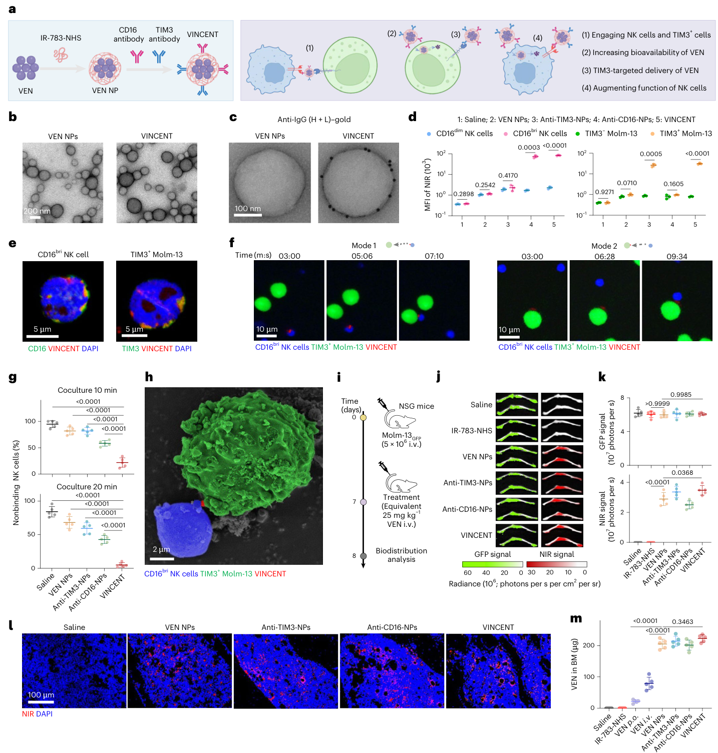
Fig. 1. Construction of VINCENT and the evaluation of targeted affinity and biodistribution in AML mice. a, Scheme of the generation of VEN NPs and loading of anti-CD16 and anti-TIM3 onto VEN NPs (VINCENT; left), along with the proposed mechanism for VINCENT (right). Scheme created in BioRender; Li, J. https://BioRender.com/5l29djy (2025). b, TEM images of VEN NPs and VINCENT. c, TEM images of immunogold analysis of VEN NPs and VINCENT using anti-IgG (H + L) antibody–gold bioconjugate. d, Mean fluorescence intensity analysis of IR-783 at NIR channel in NK cells or Molm-13 cells. e, Fluorescence visualization of VINCENT after 20-min incubation with CD16bri NK cells or TIM3+ Molm-13 cells (VINCENT, red; nuclei, blue; CD16 or TIM3, green). f, Fluorescence live imaging of VINCENT-mediated engagement between CD16bri NK cells and TIM3+ Molm-13 cells (VINCENT, red; CD16bri NK cells, blue; TIM3+ Molm-13 cells, green). g, Proportion of NK cells that do not bind to Molm-13 cells in the live imaging cocultures. Each point represents the mean proportion of nonbinding NK cells(图注取自PDF文本层,来源:Nature Cancer, 2026)
2. TIM3靶向递送能克服VEN耐药吗?→ 能,且关键在“精准”
他们先用VEN浓度梯度爬升法构建了Molm-13VEN耐药株,发现TIM3表达升高,且Lin−CD34+CD38−CD99+TIM3+ LSC-like亚群显著富集。用抗TIM3-NPs处理,IC50较游离VEN降低了一个数量级。
更关键的是:他们用UPLC-MS/MS直接定量细胞内VEN浓度。抗TIM3-NPs让TIM3+细胞内的VEN含量提高了12.8倍,而TIM3−细胞几乎没变化。同样是高浓度VEN,TIM3+细胞凋亡率却显著低于TIM3−细胞——说明TIM3本身与VEN耐药机制相关(文中未深究具体通路,但提到可能与LSC特征有关)。
@方法论点评:这里用了“药物含量定量+凋亡率”双指标交叉验证:如果单纯看杀伤效果,可能归因于靶向递送;但通过比较TIM3+和TIM3−细胞在同等胞内VEN浓度下的凋亡差异,直接证明了TIM3+细胞确实是“耐药核心人群”,靶向递送等于“绕过”了其固有耐药机制。
体内:抗TIM3-NPs在NSG小鼠中显著降低骨髓白血病负荷,延长生存至70天,优于“抗TIM3抗体+VEN NPs”的物理混合组合。
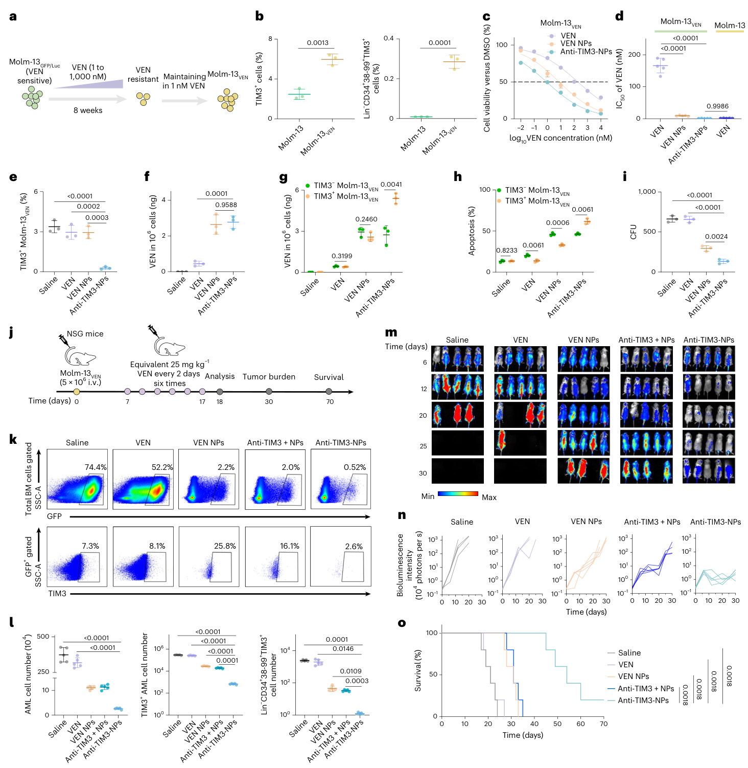
Fig. 2. Anti-TIM3-NPs enhance cellular uptake and overcome VEN resistance of AML cells. a, Schematic of Molm-13VEN construction. Cells were cultured in medium supplemented with increasing concentrations of VEN from 1 to 1,000 nM for more than 8 weeks and resistant cells were maintained in the presence of VEN. b, Percentage of TIM3+ and Lin−CD34+CD38−CD99+TIM3+ subsets in parental Molm-13 or Molm-13VEN. c, Survival of Molm-13VEN treated with different concentrations of free VEN, VEN NPs and anti-TIM3-NPs for 24 h. d, IC50 of free VEN, VEN NPs and anti-TIM3-NPs in Molm-13VEN, as well as IC50 of free VEN in Molm-13. e, Percentage of TIM3+ cells in Molm-13VEN treated with equivalent 5 nM VEN for 24 h. f,g, Quantification of VEN content in total Molm-13VEN (f) and TIM3− or TIM3+ cells (g) treated with equivalent 5 nM VEN for 24 h by UPLC–MS/MS. h, Apoptotic rate of TIM3− or TIM3+ Molm-13VEN treated with equivalent 5 nM VEN(图注取自PDF文本层,来源:Nature Cancer, 2026)
3. 那TIM3靶向递送会不会误伤T细胞?→ 反而改善了耗竭
这是另一个巧妙的地方。作者注意到VEN本身对活化的T细胞有off-target毒性,但他们提出:如果把VEN定向送到TIM3+的耗竭T细胞(Tex)里,是不是反而能清除“坏”T细胞,给“好”T细胞腾出空间?
他们用anti-CD3/CD28 beads刺激健康供者T细胞,诱导出TIM3+CD8+ Tex。抗TIM3-NPs处理显著减少了TIM3+CD8+、PD1+TIM3+、LAG3+TIM3+等耗竭亚群,同时中央记忆T细胞(Tcm)比例上升,IFN-γ、GZMB、TNF-α分泌增强。
@方法论点评:这是典型的“功能获益-表型验证”闭环——先看杀伤(清除TIM3+细胞),再看功能恢复(细胞因子、增殖),证明不是简单的“杀光了事”,而是重塑了整个T细胞亚群结构。
4. VINCENT如何提升NK细胞功能?→ 低E:T比下尤其明显
他们设置了1:1和1:3两种效靶比。在1:3(更接近体内实际情况)下,游离VEN或VEN NPs几乎无效,而VINCENT依然能显著杀伤Molm-13VEN,且残留的AML细胞克隆形成能力大幅下降。
机制上:VINCENT通过CD16交联激活NK细胞(p-CD3ζ和p-ZAP70/Syk升高),同时VEN的BCL-2抑制使AML细胞更易被NK细胞杀伤。两者协同,让NK细胞在“敌众我寡”的局面下依然能打。
他们构建了无药物负载的NCENT(仅双抗连接的空纳米颗粒)作为对照,结果NCENT+游离VEN只能短暂延缓肿瘤生长,无法治愈,而VINCENT在70天生存率达80%。这直接证明:靶向递送VEN + NK细胞重定向必须物理偶联在同一颗粒上才能发挥协同效应,缺一不可。
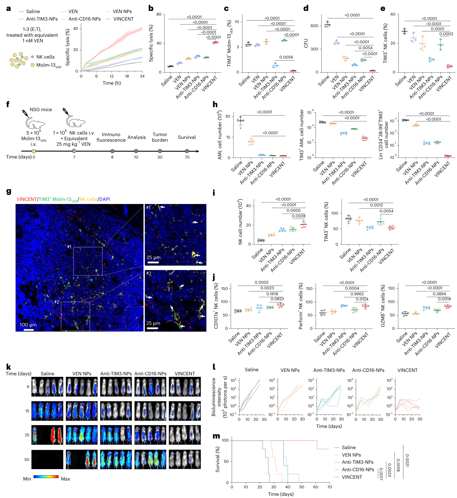
Fig. 3. VINCENT improves antileukemic response of NK cells. a, Molm-13VEN cells were used as targets in cytotoxicity assays with NK cells (E:T ratio of 1:3) by real-time measurements treated with equivalent 1 nM VEN in 24 h (Incucyte). b,c, Specific lysis of Molm-13VEN cells (b) and retained TIM3+ Molm-13VEN cell proportions (c) with NK cells (E:T ratio of 1:3) by flow cytometry measurements treated with equivalent 1 nM VEN for 24 h. d, Quantification of colonies formed by retained Molm-13VEN cells. e, Percentage of TIM3+ NK cells by flow cytometry measurements treated with equivalent 1 nM VEN for 24 h. f, Schematic of experimental design for NK cells and VINCENT combination therapy in AML cell-line-derived xenograft model. g, Fluorescence of femur BM in AML-bearing mice with VINCENT treatment (VINCENT, red; nuclei, blue; TIM3, green; CD16,(图注取自PDF文本层,来源:Nature Cancer, 2026)
5. PDX模型和患者原代样本:疗效与E:T比相关
他们在两株VEN耐药PDX模型(AML07,IC50=12.3 μM;AML10,IC50=594 nM)中验证,VINCENT均显著减少TIM3+和LSC-like亚群,提高NK细胞活化标志物(CD107a、Perforin、GZMB),延长生存。
对15例原代AML样本的分析发现:VEN耐药样本中TIM3+比例更高,且内源性NK细胞与TIM3+ blast的比例(1:3左右)远优于NK细胞与总blast的比例(1:229)。这意味着“以TIM3为锚点”的策略在临床样本中具有天然优势——不需要NK细胞很多,只要能精准识别TIM3+耐药亚群就行。
疗效改善幅度与样本IC50正相关,与NK/TIM3+ blast比例正相关,提示这是一个潜在的伴随诊断指标。
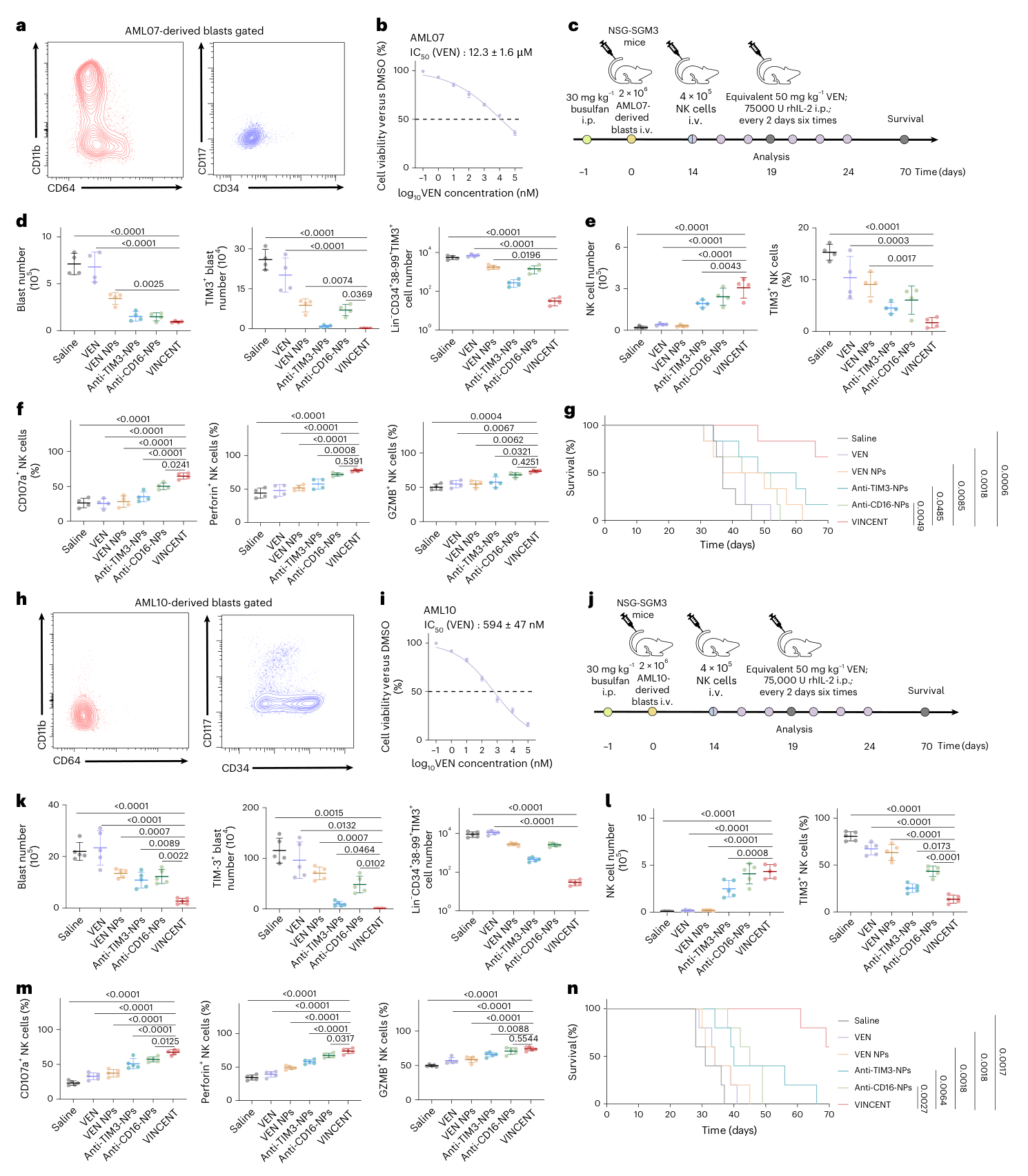
Fig. 4. VINCENT improves antileukemic effect of NK cells in AML PDX. a, The CD34/CD117 and CD64/CD11b flow plots show the immunophenotype of AML07-derived blasts. b, Viability of AML07-derived blasts treated with different concentrations of free VEN for 24 h. c, Schematic of experimental design for NK cells and VINCENT combination therapy in AML07 PDX model. d, Quantification of total, TIM3+ and Lin−CD34+CD38−CD99+TIM3+ cells in BM after treatment. e, Quantification of total NK cells and TIM3+ NK cell proportions in BM after treatment. f, Quantification of CD107a+, Perforin+ and GZMB+ NK cells proportions in BM after treatment. g, Kaplan–Meier survival curve of leukemia- bearing mice with different treatments. h, The CD34/CD117 and CD64/CD11b flow plots show the immunophenotype of AML10-derived blasts. i, Viability of AML10-derived blasts treated with different concentrations of free VEN for 24 h.(图注取自PDF文本层,来源:Nature Cancer, 2026)
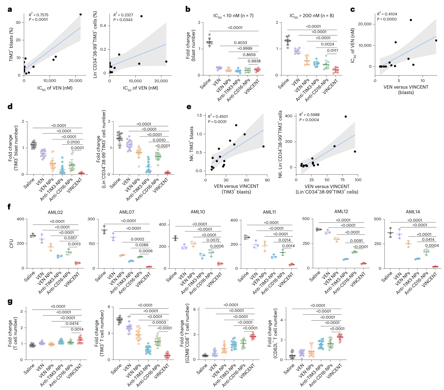
Fig. 5. VINCENT boosts endogenous NK and T cells in individual-derived samples. a, Pearson correlation between the IC50 of blasts and the percentage of TIM3+ blasts or Lin−CD34+CD38−CD99+TIM3+ cells. Pearson coefficients (R2) are provided. b, Fold change of total blasts number treated with equivalent 5 nM VEN for 24 h in VEN-sensitive (n = 7 individual samples) or VEN-resistant (n = 8 individual samples) samples. c, Pearson correlation between the improvement of VINCENT to free VEN in number of total blasts and the IC50 of blasts. Pearson coefficients are provided. d, Fold change of TIM3+ blasts and Lin−CD34+CD38−(图注取自PDF文本层,来源:Nature Cancer, 2026)
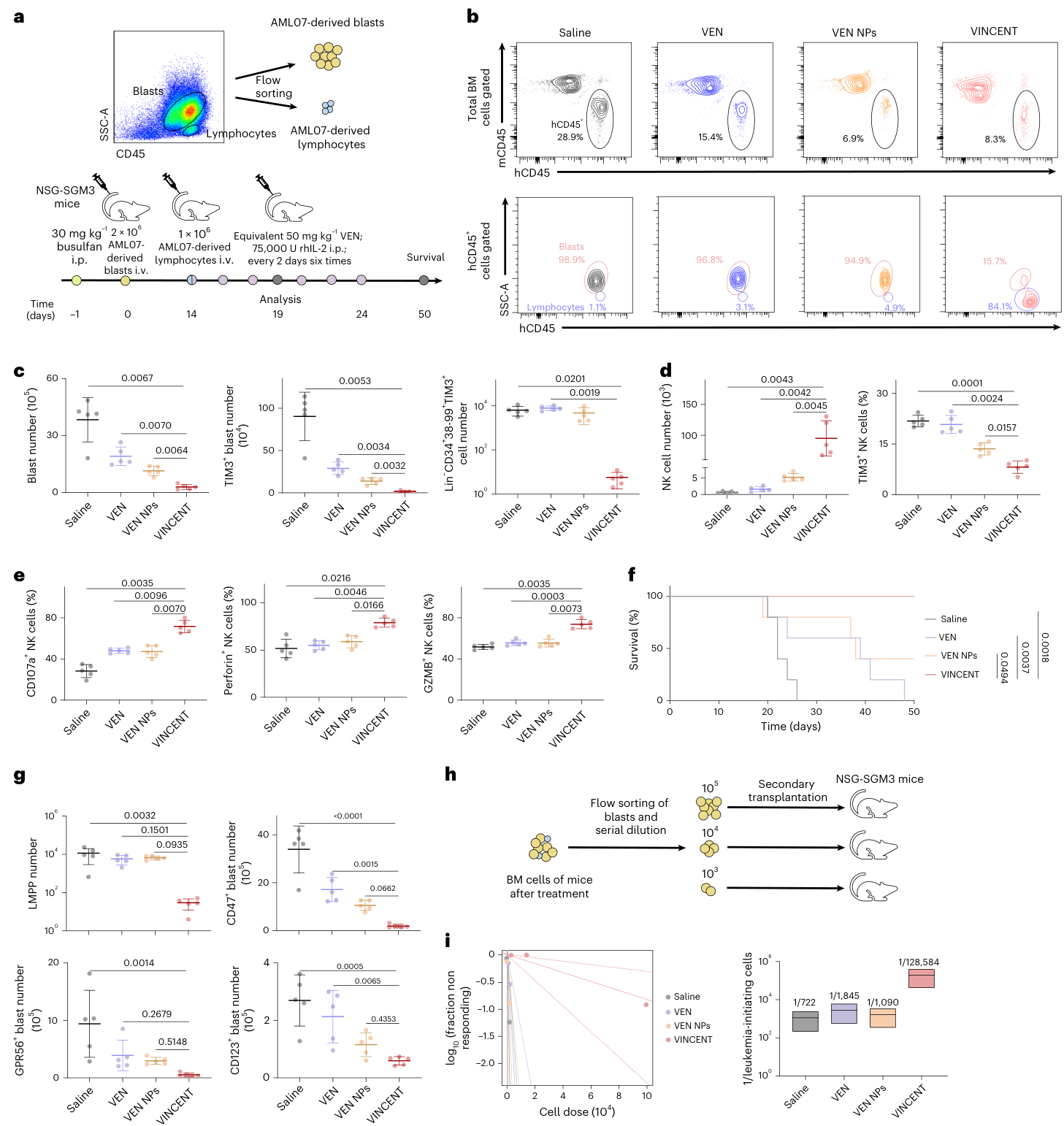
Fig. 6. VINCENT reinvigorates endogenous NK cell activity in AML PDX. a, Schematic of experimental design for individual-derived lymphocytes and VINCENT combination therapy in PDX model. b, Representative flow plots of BM cell analysis. c, Quantification of total, TIM3+ and Lin−CD34+CD38−CD99+TIM3+ cells in BM after treatment. d, Quantification of total NK cells and TIM3+ NK cell proportions in BM after treatment. e, Quantification of CD107a+, Perforin+ and GZMB+ NK cell proportions in BM after treatment. f, Kaplan–Meier survival curve of leukemia-bearing mice with different treatment. g, Quantification of LMPPs, CD47+, GPR56+ and CD123+ blasts in BM after treatment. Data were derived(图注取自PDF文本层,来源:Nature Cancer, 2026)
6. 单细胞测序揭示:VINCENT重塑了整个骨髓微环境
scRNA-seq显示,VINCENT治疗后AML细胞显著减少,且高表达HAVCR2(TIM3编码基因)的亚群被选择性清除。NK细胞增多且PRF1、GZMB等杀伤基因上调。单核细胞中CD16a+亚群增加,提示可能激活了炎症性单核细胞,进而促进T细胞应答。
T细胞层面:GZMB+CD8+ CTL比例上升,TIM3+PD1+共表达耗竭亚群比例下降。细胞通讯分析显示VINCENT组中NK-单核-T细胞之间的配体-受体交互增强,说明它不是一个“单打独斗”的药,而是“搅动”了整个免疫网络。
@方法论点评:scRNA-seq的价值在于看到了“出乎意料的效应”——VINCENT不仅清除了目标细胞,还改变了单核细胞亚群分布和细胞间通讯网络,为后续研究提供了新方向。
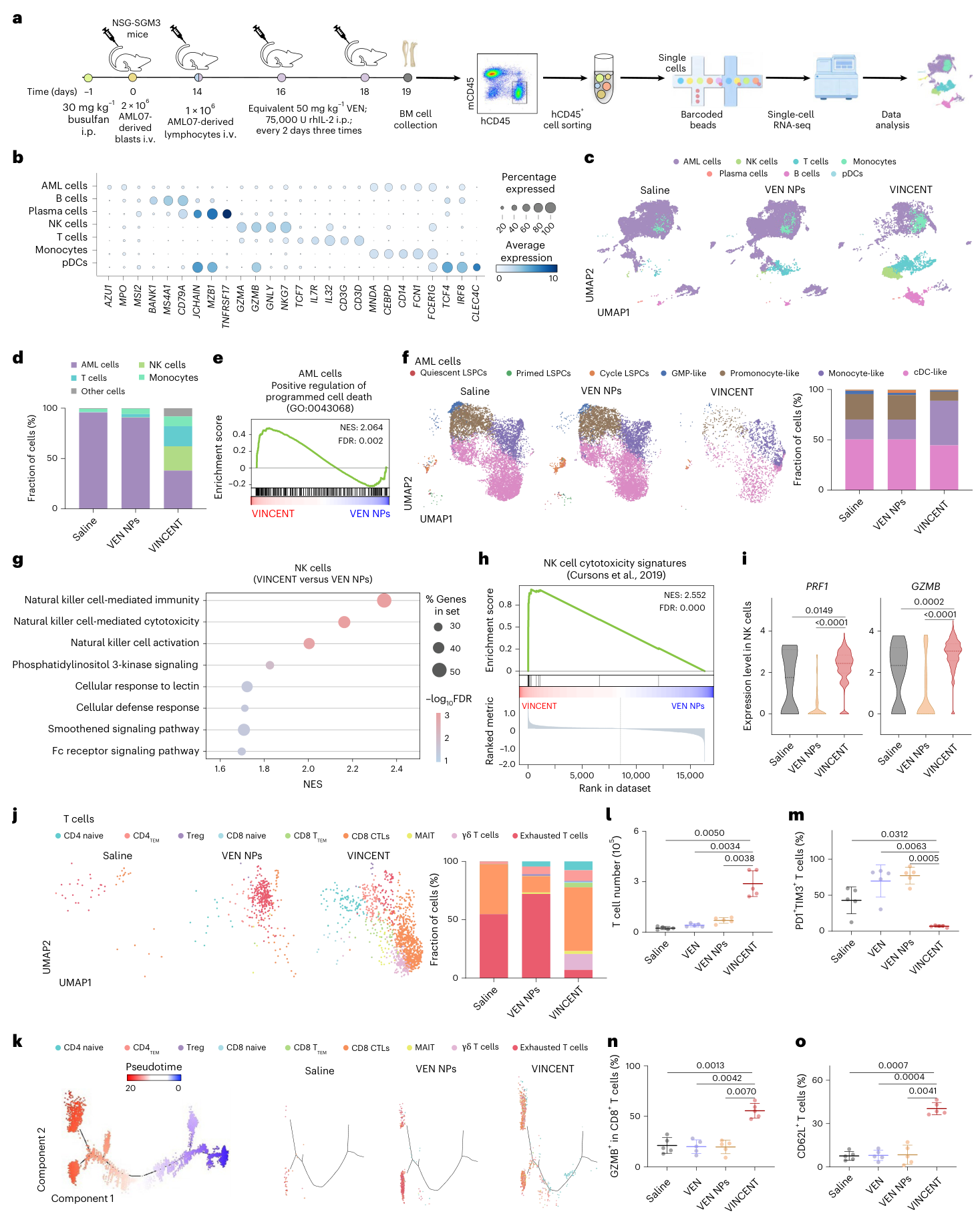
Fig. 7. scRNA-seq reveals VINCENT reshapes the immunosuppressive leukemic microenvironment. a, Schematic for generating scRNA-seq. Human CD45+ cells were sorted and pooled from BM cells of mice with individual-derived lymphocytes and VEN NPs/VINCENT combination therapy in PDX model (n = 5 mice). b, A dot plot displaying conserved marker genes in each cell population. c, UMAP plot of scRNA-seq data from human CD45+ cells with seven cell clusters annotated. d, The fraction of AML cells, NK cells, T cells, monocytes and other cell populations among total human CD45+ cells. e, GSEA enrichment plots of genes associated with positive regulation of programmed cell death enriched in AML cells. NES, normalized enrichment score; FDR, false discovery rate. f, UMAP plot of scRNA-seq data from AML cells with seven cell clusters annotated (left) and the fraction of each AML subpopulation (right). g, Gene Ontology analysis of(图注取自PDF文本层,来源:Nature Cancer, 2026)
核心结论
VINCENT集成了三项功能:①靶向递送VEN到TIM3+耐药细胞;②通过CD16激活内源性NK细胞;③清除耗竭T细胞,改善免疫微环境。它在耐药细胞系、PDX模型和患者原代样本中均展示了协同抗白血病效应,且安全性良好(不影响正常HSC和主要脏器)。
对耐药/DTP/PGCC 的启示
靶向“耐药亚群表面标志物”进行药物递送:TIM3+细胞不仅是耐药主体,还是DTP的潜在“庇护所”。VINCENT的思路可推广到其他DTP标志物(如CD47、GPR56),实现“标志物导向的化疗增敏”。 免疫微环境重编程是克服DTP持久性的关键:DTP/PGCC常通过免疫逃逸存活;VINCENT同时清除耐药细胞和唤醒NK/T细胞,相当于“拆了DTP的围墙”。未来可探索与免疫检查点抑制剂(如抗PD-1)的序贯或联合策略。 E:T比可作为临床富集标志物:文中明确提出“NK细胞/TIM3+ blast比例”可预测VINCENT疗效,这种“靶向细胞 vs 效应细胞”的比值思维,对设计DTP靶向免疫疗法具有量化指导意义。
局限
TIM3与VEN耐药的具体分子机制未阐明(文中仅提示与LSC特征相关,未深入BCL-2家族或代谢通路变化)。 NSG/NSG-SGM3小鼠缺乏完整的适应性免疫系统,人源免疫细胞重建后的长期免疫记忆形成未评估。 纳米颗粒的规模化生产和长期稳定性、临床给药方案(剂量、频次)尚未讨论。 安全性数据仅在健康小鼠中评估,未在荷瘤长期存活小鼠中系统评估对正常HSC的累积影响。
来源
期刊:Nature Cancer,2026。DOI: 10.1038/s43018-026-01217-z