一句话亮点
这项研究发现,棕榈酰转移酶ZDHHC16通过催化GPX2蛋白C67位点的S-棕榈酰化,阻止其被E3泛素连接酶SYVN1降解,从而稳定GPX2蛋白水平,抑制乐伐替尼诱导的免疫原性细胞死亡(ICD)和抗肿瘤免疫,最终推动肝癌乐伐替尼耐药。靶向这一棕榈酰化修饰可恢复乐伐替尼的疗效。
背景/痛点
乐伐替尼是晚期肝癌的一线靶向药,但耐药问题严重制约了其临床获益。虽然已知耐药机制涉及信号通路异常、代谢重编程、微环境改变等多个层面,但对蛋白质翻译后修饰如何调控耐药,尤其是S-棕榈酰化这种可逆脂质修饰在其中的角色,认知还很有限。
另一方面,乐伐替尼除了直接抑制肿瘤细胞增殖外,还能诱导ICD——一种"有组织有预谋"的细胞死亡方式,释放DAMPs(如CRT、HMGB1、ATP),召集合抗原呈递细胞,激活T细胞抗肿瘤免疫。问题是:肿瘤细胞如何逃避乐伐替尼诱导的ICD?这个问题的答案,可能正是耐药的突破口。
GPX2此前已被报道与肝癌乐伐替尼耐药相关,但调控其稳定性的上游机制尚不清楚。作者团队决定从棕榈酰转移酶家族入手,系统性筛选与乐伐替尼耐药相关的关键酰基转移酶。
推理链分步拆解
第一步:大海捞针——谁在调控乐伐替尼敏感性?
作者首先在已建立的乐伐替尼耐药肝癌细胞株中,逐一敲低了23个棕榈酰转移酶家族成员,观察哪个基因的沉默能最大程度地降低乐伐替尼的IC50。结果ZDHHC16脱颖而出——敲低它带来的增效最显著。
耐药株中ZDHHC16的蛋白水平明显高于亲本株;在临床肝癌组织样本中,乐伐替尼耐药组患者的肿瘤组织ZDHHC16表达也显著高于敏感组。TCGA数据库分析还显示,ZDHHC16高表达与更差的无病生存和总生存相关。
这些结果将ZDHHC16推到了聚光灯下,但截至目前仍然是相关性证据——ZDHHC16高表达伴随耐药,还不能证明它有功能性贡献。
@方法论点评:在23个同家族成员中进行功能性筛选,是典型的“先宽后窄”策略,避免先验假设带来的偏见。IC50作为初筛指标合理,但后续必须用遗传敲除和药物联合实验来确认因果关系。
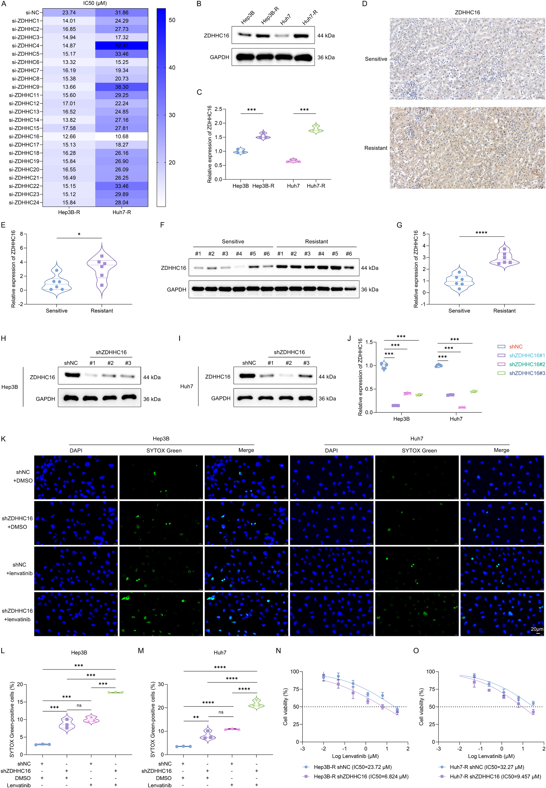
Fig. 1. ZDHHC16-mediated S-palmitoylation is crucial for regulating HCC sensitivity to lenvatinib. (A) Heatmap showing the IC50 value of lenvatinib in different groups. Hep3B-R and Huh7-R cells were transfected with specific siRNAs of 23 palmitoyl acyltransferases and corresponding controls (si-NC) for 24 h, and treated with a series of doses of lenvatinib for 24 h. Cell viability was detected through CCK−8 assay, and the IC50 value of lenvatinib was calculated. (B, C) Western blot for the expression of ZDHHC16 in lenvatinib-resistant Hep3B and Huh7 cells (Hep3B-R and Huh7-R) and their parental cells. (D, E) IHC staining for detecting the expression of ZDHHC16 in lenvatinib-sensitive and -resistant tumor tissues derived from HCC patients (n = 6 per group). Scale bar, 50 μm. (F, G) Western blot for detecting the expression of ZDHHC16 in lenvatinib-sensitive and -resistant tumor tissues derived from HCC patients (n = 6 per group). (H-J) Western blot for the expression of ZDHHC16 in Hep3B and Huh7 cells that were transfected with three ZDHHC16 shRNAs (shZDHHC16#1, #2, #3) and corresponding controls (shNC) for 48 h. (K-M) SYTOX Green for analyzing the dead cells in Hep3B and Huh7 cells that were transfected with shZDHHC16 or shNC for 48 h and treated with 10 μM lenvatinib or DMSO for 24 h. Scale bar, 20 μm. (N, O) The IC50 value of lenvatinib in lenvatinib-resistant HCC cells. Hep3B-R and Huh7-R cells were transfected with shZDHHC16 or shNC for 48 h, followed by treatment with a series of lenvatinib doses for 24 h. After measuring cell viability using CCK−8 assay, the IC50 value of lenvatinib was calculated. , P < 0.05; , P < 0.01; , P < 0.001; , P < 0.0001; ns, P > 0.05 from Student’s t test or one-way ANOVA followed by Tukey’s post hoc test.(图注取自PDF文本层,来源:Drug Resistance Updates, 2026)
第二步:ZDHHC16沉默如何改变了细胞死亡的“性质”?
既然ZDHHC16与耐药相关,敲低它会怎样改变细胞对乐伐替尼的响应?
作者发现ZDHHC16敲低本身就能轻微增加肿瘤细胞死亡,但与乐伐替尼联用后,细胞死亡显著加剧——而且不是普通的凋亡。他们检测了ICD的核心标志物:CRT膜转位(“吃我”信号)、HMGB1和ANXA1释放、ATP释放。
单用乐伐替尼能诱导一定程度的CRT转位和DAMP释放,但力度有限。而ZDHHC16敲低+乐伐替尼,则让这些指标大幅飙升。同时,细胞内ROS水平急剧升高,ER stress标志物(p-PERK、p-IRE1α、ATF6、CHOP)上调。敲低ZDHHC16和乐伐替尼在诱导ER stress上有明确的协同效应。
这组实验清楚地指向一个结论:ZDHHC16天然地抑制乐伐替尼诱导的ICD,敲除它等于“松开刹车”。
@方法论点评:这里的关键是区分“细胞死亡”和“免疫原性细胞死亡”。作者没有只看细胞死活,而是系统检测了ICD的核心DAMP释放谱,这是验证ICD性质的行业金标准。ER stress和ROS的检测则为后续机制埋下伏笔。
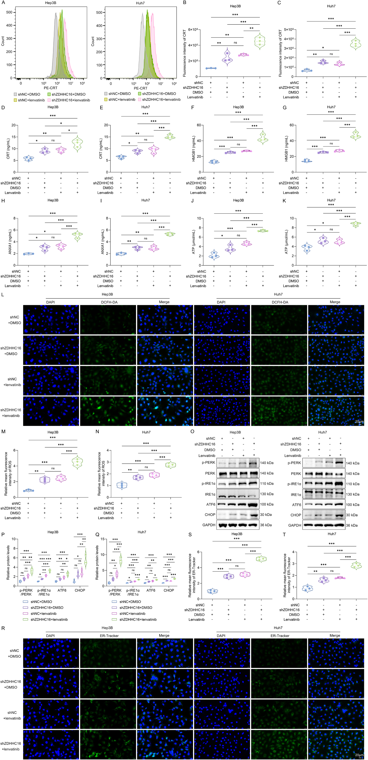
Fig. 2. Knockdown of ZDHHC16 improves lenvatinib-induced ICD in HCC cells. (A) Representative histogram illustrating the percentage of CRT-expressing Hep3B and Huh7 cells as measured by flow cytometry. Cells were transfected with shZDHHC16 or shNC for 48 h and treated with 10 μM lenvatinib or DMSO for 24 h. (B, C) Mean fluorescence intensity value for CRT staining. (D-K) ELISA assays for measuring the extracellular levels of (D, E) CRT, (F, G) HMGB1, (H, I) ANXA1, and (J, K) ATP in the supernatant of HCC cells that were transfected with shZDHHC16 or shNC for 48 h and treated with 10 μM lenvatinib or DMSO for 24 h. (L-N) DCFH-DA probe staining for detecting the intracellular ROS levels in HCC cells with transfection of shZDHHC16 or shNC for 48 h and treatment with 10 μM lenvatinib or DMSO for 24 h. Scale bar, 20 μm. (O-Q) Western blot for measuring the expression levels of phosphorylated PERK and IRE1α, as well as ATF6 and CHOP in HCC cells, after transfecting with shZDHHC16 or shNC for 48 h and treating with 10 μM lenvatinib or DMSO for 24 h. (R-T) ER-Tracker Green fluorescent probe staining for evaluation of ER stress in HCC cells with transfection of shZDHHC16 or shNC for 48 h and treatment with 10 μM lenvatinib or DMSO for 24 h. Scale bar, 20 μm. , P < 0.05; , P < 0.01; , P < 0.001; ns, P > 0.05 from one-way ANOVA followed by Tukey’s post hoc test.(图注取自PDF文本层,来源:Drug Resistance Updates, 2026)
第三步:在动物身上复现——免疫系统参与进来
体外数据再漂亮,也得在免疫完整的动物模型里验证。作者将ZDHHC16敲低或对照的Hepa1-6小鼠肝癌细胞皮下接种到C57BL/6小鼠体内,待成瘤后给予乐伐替尼口服治疗。
结果与体外高度一致:ZDHHC16敲低+乐伐替尼组肿瘤生长最慢、重量最轻,CRT表达和DAMP释放最强,ROS积累最显著。更重要的是,肿瘤免疫微环境发生了决定性转变——CD8⁺T细胞、成熟DC、M1型巨噬细胞浸润增加,而免疫抑制性的Treg和M2型巨噬细胞减少。
这说明ZDHHC16敲低不仅直接增强了肿瘤细胞的ICD,还重塑了免疫微环境,使之从“冷”转“热”,放大了乐伐替尼的免疫协同效应。
@方法论点评:体内实验是验证ICD-抗肿瘤免疫轴不可或缺的一环。体外看到DAMP释放还不够,体内必须看到T细胞浸润和免疫微环境重塑才算闭环。作者采用了免疫健全的C57BL/6模型而非裸鼠,保留了适应性免疫系统,这比免疫缺陷模型更有说服力。

Fig. 3. Blockade of ZDHHC16 combined with lenvatinib inhibits tumor growth through enhancing ICD-elicited antitumor immunity. (A) Schematic diagram of the experimental design. Hepa 1–6 cells with shNC and shZDHHC16 were subcutaneously injected into the mice. When tumors reached 200 mm³ , the mice were administered with either lenvatinib (10 mg/kg/day, orally) or DMSO. On the 19th day after subcutaneous injection, the tumors were gathered and measured. (B) Body weight changes of C57BL/6 J mice. (C, D) Photographs of tumor-bearing C57BL/6 J mice and tumors. (E) Tumor growth curve. The tumor volume was measured every two days. (F) Tumor weight. (G, H) IHC staining of CRT in tumors. Scale bar, 20 μm. (I-N) ELISA assays for measuring the levels of (I) CRT, (J) HMGB1, (K) ANXA1, (L) ATP, (M) TNF-α, and (N) IFN-γ in tumors. (O, P) DCFH-DA probe staining for detecting ROS levels in tumors. Scale bar, 20 μm. (Q, R) Flow cytometry analysis of the ratios of CD8+ T cells, regulatory T cells (Tregs), mature dendritic cells (DCs), M1 macrophages, and M2 macrophages in tumors. , P < 0.05; , P < 0.01; , P < 0.001; , P < 0.0001; ns, P > 0.05 from one- or two-way ANOVA followed by Tukey’s post hoc test.(图注取自PDF文本层,来源:Drug Resistance Updates, 2026)
第四步:GPX2是如何被“粘”在细胞里的?
机制深挖的核心问题是:ZDHHC16作为一个棕榈酰转移酶,它的底物是谁?
作者通过SWISS PALM数据库预测,并结合前期文献,将目标锁定在GPX2——一个已知与乐伐替尼耐药相关的抗氧化酶。敲低ZDHHC16后,GPX2的mRNA水平不变,蛋白水平却显著下降。这提示ZDHHC16在翻译后水平调控GPX2。
接下来的棕榈酰化实验(ABE和Click-iT)证实GPX2确实被棕榈酰化,且这种修饰依赖ZDHHC16。通过构建GPX2四个半胱氨酸位点的突变体,他们精确锁定了C67是关键的棕榈酰化位点——只有C67S突变会大幅降低GPX2的棕榈酰化水平,且ZDHHC16过表达无法恢复。CHX放线菌酮追踪实验表明,C67S突变显著缩短了GPX2的蛋白半衰期。
那么这里有个问题:棕榈酰化如何延长蛋白寿命?通常有三种可能:改变蛋白构象、影响蛋白-蛋白互作、或阻断降解信号。作者排除了构象和定位的猜测,转向降解通路。
@方法论点评:一步一步卡位——“mRNA不变但蛋白变”提示翻译后修饰;“点突变锁定C67”明确了修饰位点;“半衰期缩短”直接证明稳定性变化。这三个证据链环环相扣,是经典的修饰-功能验证策略。

Fig. 4. ZDHHC16 enhances the protein stability of GPX2 by catalyzing the S-palmitoylation of GPX2 at C67 residue. (A, B) qRT-PCR for the mRNA levels of ZDHHC16 and GPX2 in HCC cells transfected with shNC or shZDHHC16 lentiviral vectors. (C-F) Western blot of the protein levels of ZDHHC16 and GPX2 in HCC cells transfected with shNC or shZDHHC16. (G) IF confocal microscopy showing the colocalization of ZDHHC16 and GPX2 in lenvatinib-resistant HCC cells and their parental cells that were transfected with shNC or shZDHHC16. Scale bar, 10 μm. (H, I) Quantification of Pearson’s colocalization coefficient (PCC) of ZDHHC16 and GPX2. (J) Western blot of GPX2 S-palmitoylation by ABE assay following treatment with DMSO or 2-BP (10 μM) for 12 h in HCC cells. (K) Western blot of GPX2 S- palmitoylation by Click-iT reaction assay following treatment with palmitic acid-azide (50 μM) with or without 2-BP (10 μM) for 8 h. (L) Western blot of GPX2 S- palmitoylation by ABE assay in HCC cells transfected with shNC or shZDHHC16. (M) Western blot of GPX2 S-palmitoylation by ABE assay in HEK293T cells transfected with plasmids encoding wild-type (WT) GPX2-HA or its indicated mutants. (N) Western blot of GPX2 S-palmitoylation by ABE assay in HEK293T cells co- transfected with plasmids encoding ZDHHC16-Flag and GPX2-WT-HA or GPX2-C67S-HA. (O-Q) Western blot of GPX2 in HCC cells transfected with GPX2-WT or GPX2-C67S, followed by CHX treatment for the indicated duration. (R-T) Western blot of GPX2 in HCC cells transfected with shNC or shZDHHC16, followed by CHX treatment for the indicated duration. , P < 0.05; , P < 0.01; , P < 0.001; , P < 0.0001; ns, P > 0.05 from one- or two-way ANOVA followed by Tukey’s post hoc test.(图注取自PDF文本层,来源:Drug Resistance Updates, 2026)
第五步:棕榈酰化如何“保护”GPX2不被降解?
既然棕榈酰化稳定了GPX2,那它稳定GPX2的机制是什么?
作者用MG132(蛋白酶体抑制剂)处理细胞后发现,原本因ZDHHC16敲低或C67S突变而下降的GPX2蛋白水平被“救回”了——说明GPX2是通过泛素-蛋白酶体途径降解的。泛素化实验进一步证实:ZDHHC16过表达降低GPX2泛素化水平,而这个效应在C67S突变体中消失。
那谁负责给GPX2打上泛素标签?数据库预测指向SYVN1(一种E3泛素连接酶)。Co-IP验证了SYVN1和GPX2确实存在相互作用。功能实验显示:SYVN1过表达增加GPX2泛素化并降低其蛋白水平;而SYVN1沉默则相反。重要的是,ZDHHC16表达水平能反向调节SYVN1对GPX2的泛素化效率。
综合起来,机制图像逐渐清晰:ZDHHC16将16碳棕榈酰链共价连接到GPX2的C67位点,这个脂质修饰要么从空间上挡住了SYVN1的结合位点,要么改变了GPX2的构象使其不易被识别,从而抑制了GPX2的泛素化降解。
@方法论点评:这是整个故事最精彩的部分——揭示了棕榈酰化和泛素化两种翻译后修饰之间的“对话”。棕榈酰化通过“抢占”或“遮蔽”阻止泛素化,这是蛋白质稳定性调控的一种精巧策略。
第六步:因果链闭环——GPX2棕榈酰化是ZDHHC16功能的“命门”
前面的机制已经阐明ZDHHC16→GPX2 C67棕榈酰化→稳定GPX2这条路径。但最关键的因果验证还没做:ZDHHC16对乐伐替尼ICD的抑制作用,到底是不是完全依赖GPX2 C67棕榈酰化?
作者设计了一个经典的“回复实验”:在敲低ZDHHC16的同时,分别回补野生型GPX2(GPX2-WT)或棕榈酰化缺陷突变体GPX2-C67S,然后观察ICD指标。
结果非常漂亮:在GPX2-WT细胞中,ZDHHC16敲低能够增强CRT转位、DAMP释放、ROS积累和ER stress;但在GPX2-C67S细胞中,ZDHHC16敲低的所有这些效果都被“抹平”了。耐药细胞中的IC50实验也得到同样模式——GPX2-C67S使细胞对乐伐替尼的敏感性不再受ZDHHC16调控。
@方法论点评:这就是所谓的“遗传挽救实验”——要证明A通过B发挥作用,最好的办法是让B“失效”(突变),看A的功能是否随之消失。本实验中,C67S突变完美地“截断”了ZDHHC16敲低带来的所有表型,有力地证明了因果关系的方向性和特异性。这是整个故事逻辑链条中最关键的一颗铆钉。
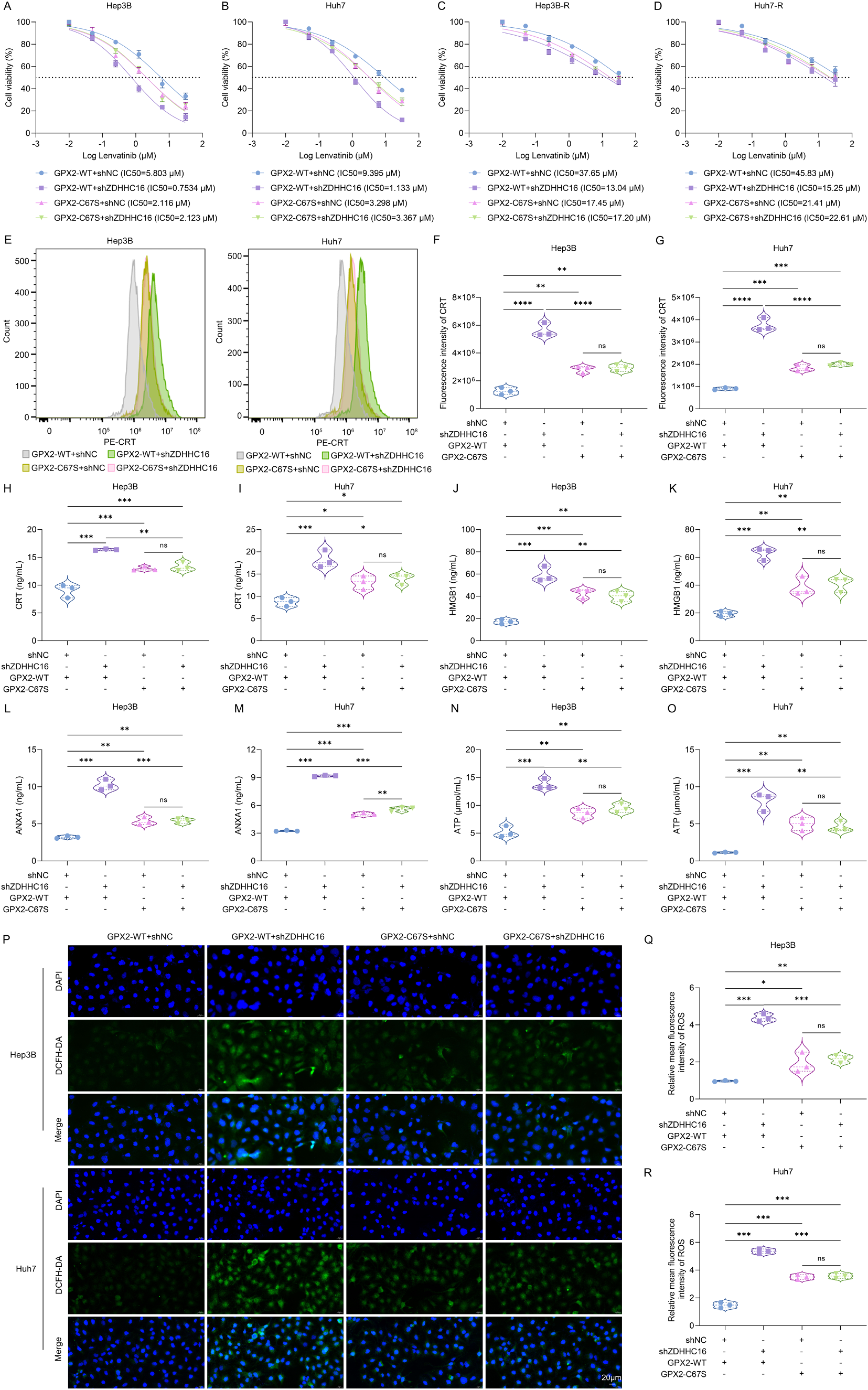
Fig. 6. ZDHHC16 inhibits lenvatinib-induced ICD by catalyzing the S-palmitoylation of GPX2. (A-D) The IC50 value of lenvatinib in Hep3B, Huh7, Hep3B-R, and Huh7-R cells that were co-transfected with shZDHHC16/shNC and GPX2-WT/GPX2-C67S, followed by treatment with different doses of lenvatinib. (E-G) Flow cytometry analysis of the percentage of CRT-expressing cells in Hep3B and Huh7 cells that were co-transfected with shZDHHC16/shNC and GPX2-WT/GPX2-C67S as well as treated with DMSO or lenvatinib (10 μM). (H-O) ELISA assays for measuring the extracellular levels of (H, I) CRT, (J, K) HMGB1, (L, M) ANXA1, and (N, O) ATP in the supernatant of HCC cells that were co-transfected with shZDHHC16/shNC and GPX2-WT/GPX2-C67S as well as treated with DMSO or lenvatinib (10 μM). (P-R) DCFH-DA probe staining for detecting the intracellular ROS levels in HCC cells that were co-transfected with shZDHHC16/shNC and GPX2-WT/GPX2-C67S as well as treated with DMSO or lenvatinib (10 μM). Scale bar, 20 μm. , P < 0.05; , P < 0.01; , P < 0.001; , P < 0.0001; ns, P > 0.05 from one-way ANOVA followed by Tukey’s post hoc test.(图注取自PDF文本层,来源:Drug Resistance Updates, 2026)
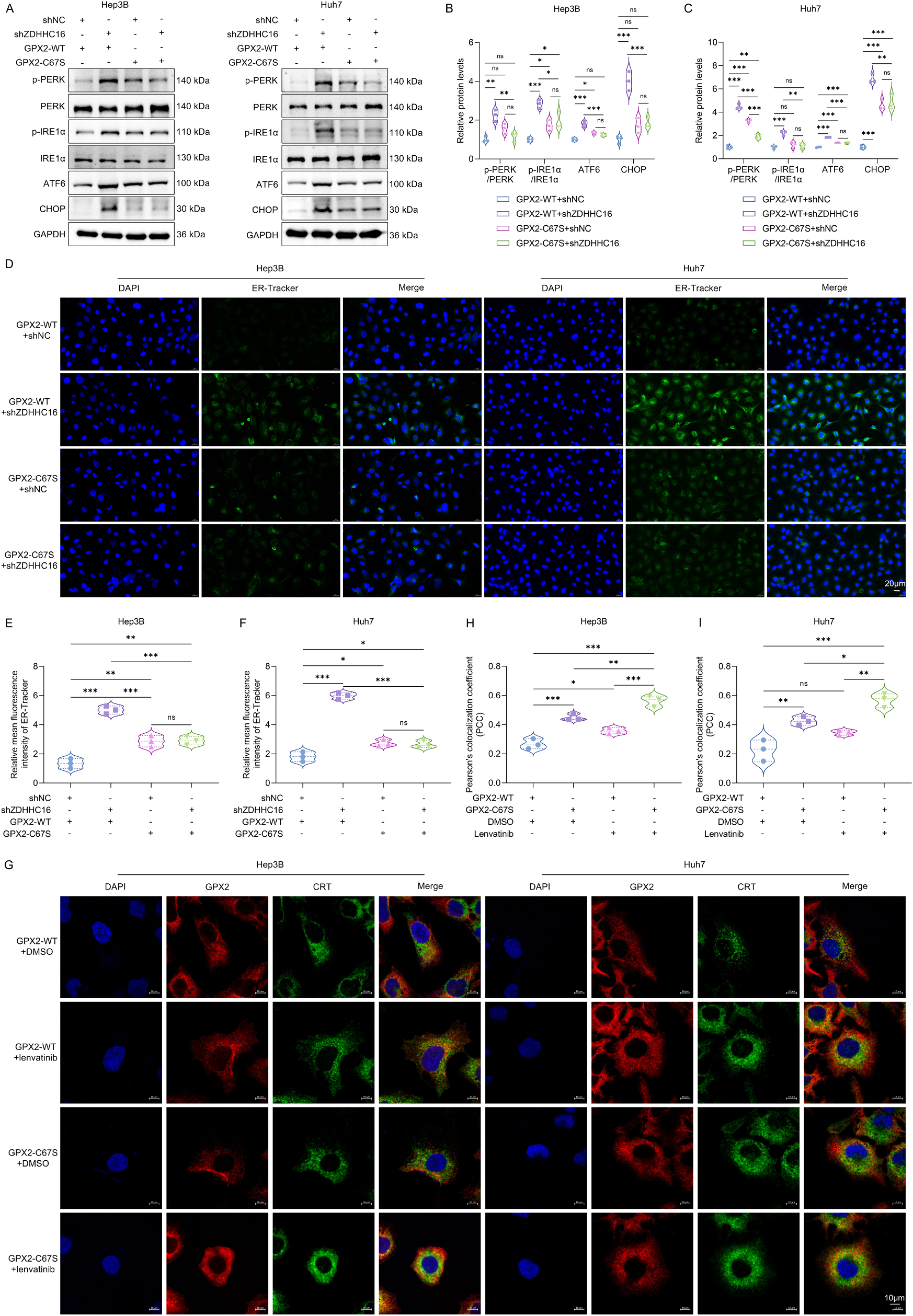
Fig. 7. ZDHHC16 inhibits lenvatinib-induced ER stress in HCC cells by catalyzing the S-palmitoylation of GPX2. (A-C) Western blot for measuring the expression levels of phosphorylated PERK and IRE1α as well as ATF6 and CHOP in Hep3B and Huh7 cells that were co-transfected with shZDHHC16/shNC and GPX2-WT/GPX2- C67S as well as treated with DMSO or lenvatinib (10 μM). (D-F) ER-Tracker Green fluorescent probe staining for evaluation of ER stress in HCC cells that were co- transfected with shZDHHC16/shNC and GPX2-WT/GPX2-C67S as well as treated with DMSO or lenvatinib (10 μM). Scale bar, 20 μm. (G) IF confocal microscopy showing the colocalization of GPX2 and CRT in HCC cells that were transfected with GPX2-WT/GPX2-C67S and treated with DMSO or lenvatinib (10 μM). Scale bar, 10 μm. (H, I) Quantification of Pearson’s colocalization coefficient (PCC) of GPX2 and CRT. , P < 0.05; , P < 0.01; , P < 0.001; ns, P > 0.05 from one-way ANOVA followed by Tukey’s post hoc test.(图注取自PDF文本层,来源:Drug Resistance Updates, 2026)

Fig. 8. ZDHHC16-mediated GPX2 S-palmitoylation induces ICD in HCC cells by inducing ER stress. (A-C) Flow cytometry analysis of the percentage of CRT- expressing cells in shNC/shZDHHC16 and GPX2-WT/GPX2-C67S HCC cells that were treated with 1 mM 4-PBA for 24 h. (D-K) ELISA assays for measuring the extracellular levels of (D, E) CRT, (F, G) HMGB1, (H, I) ANXA1, and (J, K) ATP in the supernatant of shNC/shZDHHC16 and GPX2-WT/GPX2-C67S HCC cells that were treated with 1 mM 4-PBA for 24 h. (L-N) DCFH-DA probe staining for detecting the intracellular ROS levels in shNC/shZDHHC16 and GPX2-WT/GPX2-C67S HCC cells that were treated with 1 mM 4-PBA for 24 h. Scale bar, 20 μm. (O-Q) ER-Tracker Green fluorescent probe staining for evaluation of ER stress in shNC/ shZDHHC16 and GPX2-WT/GPX2-C67S HCC cells that were treated with 1 mM 4-PBA for 24 h. Scale bar, 20 μm. , P < 0.01; , P < 0.001; , P < 0.0001 from one-way ANOVA followed by Tukey’s post hoc test.(图注取自PDF文本层,来源:Drug Resistance Updates, 2026)
第七步:体内最后一道验证——靶向GPX2棕榈酰化真的能增敏乐伐替尼吗?
最后,作者回到动物模型,将GPX2-WT或C67S突变体与ZDHHC16敲低共转染的Hepa1-6细胞接种小鼠,然后给予乐伐替尼治疗。
结果与体外完全呼应:在GPX2-WT肿瘤中,ZDHHC16敲低显著抑制肿瘤生长、增加CRT表达和DAMP释放、促进CD8⁺T细胞浸润;但在GPX2-C67S肿瘤中,ZDHHC16敲低的所有抑瘤和免疫激活效应全部消失。
@方法论点评:从体外到体内、从细胞到动物的多重验证,极大增强了结论的外推价值。特别是体内实验中免疫微环境指标的全面评估,将分子机制与抗肿瘤免疫治疗这一临床相关维度成功对接。

Fig. 9. Suppression of ZDHHC16 enhances ICD and antitumor immunity induced by lenvatinib by GPX2 S-palmitoylation. (A) Schematic diagram of the experimental design. Hepa 1–6 cells co-transfected with shZDHHC16/shNC and GPX2-WT/GPX2-C67S were subcutaneously injected into C57BL/6 J mice. When tumors reached 200 mm³ , the mice were administrated with lenvatinib (10 mg/kg/day, orally). On the 19th day after subcutaneous injection, the tumors were excised and measured. (B) Body weight changes of C57BL/6 J mice. (C, D) Photographs of tumor-bearing C57BL/6 J mice and tumors. (E) Tumor growth curve. The tumor volume was monitored every two days. (F) Tumor weight. (G, H) IHC staining of CRT in tumors. Scale bar, 20 μm. (I) IF photographs of ZDHHC16, GPX2, and CRT in mouse tumors. Scale bar, 100 μm. (J-O) ELISA assays for measuring the levels of (J) CRT, (K) HMGB1, (L) ANXA1, (M) ATP, (N) TNF-α, and (O) IFN-γ in tumors. (P, Q) DCFH-DA probe staining for detecting ROS levels in tumors. Scale bar, 20 μm. (R, S) Flow cytometry analysis of the ratios of CD8+ T cells, regulatory T cells (Tregs), mature dendritic cells (DCs), M1 macrophages, and M2 macrophages in tumors. , P < 0.05; , P < 0.01; , P < 0.001; ns, P > 0.05 from one- or two-way ANOVA followed by Tukey’s post hoc test.(图注取自PDF文本层,来源:Drug Resistance Updates, 2026)
核心结论
这项研究揭示了一条完整的肝癌乐伐替尼耐药新通路:
ZDHHC16高表达 → GPX2 C67位点S-棕榈酰化 → 阻止SYVN1介导的泛素化降解 → GPX2蛋白稳定 → 清除乐伐替尼诱导的ROS → 抑制ER stress → 阻断ICD及抗肿瘤免疫激活 → 乐伐替尼耐药
从治疗角度,靶向这一棕榈酰化修饰(抑制ZDHHC16或破坏GPX2 C67位点)可恢复乐伐替尼诱导ICD的能力,将“冷”肿瘤转化为“热”肿瘤,协同增强乐伐替尼的疗效。
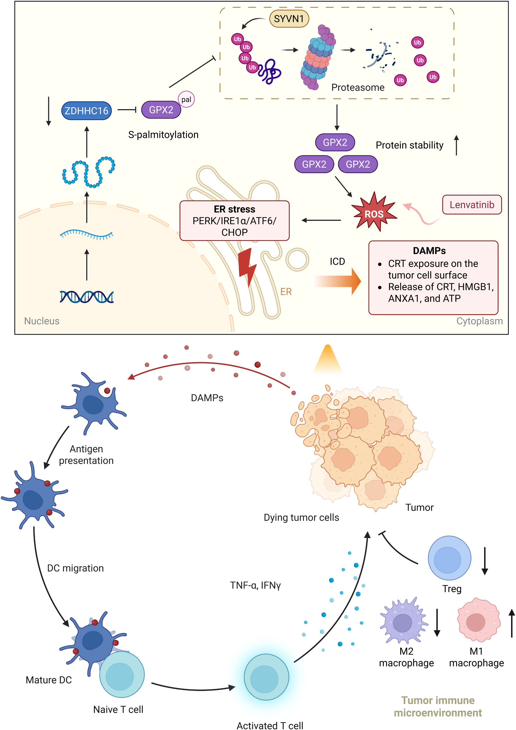
Fig. 10. A schematic diagram of this study. ZDHHC16 is responsible for catalyzing the S-palmitoylation of GPX2 at C67 and improves its protein stability via preventing ubiquitination-proteasome degradation, thereby suppressing lenvatinib-elicited ICD and antitumor immunity in HCC and contributing to lenvati nib resistance.(图注取自PDF文本层,来源:Drug Resistance Updates, 2026)
对耐药/DTP/PGCC 的启示
蛋白质翻译后修饰是耐药可塑性的“快速响应层”:相比于基因突变,棕榈酰化是动态可逆的,肿瘤细胞可能通过快速调整ZDHHC16活性来适应药物压力,形成“非遗传性耐药”。这提示在靶向治疗中同时干预修饰酶可能阻断耐药早期的“软着陆”。 ICD的“质”比“量”更关键:乐伐替尼本身就能杀死一部分肿瘤细胞,但如果死亡方式是非免疫原性的,抗肿瘤免疫就无法有效启动。本研究表明,ZDHHC16-GPX2轴可能正是决定乐伐替尼诱导的细胞死亡“是否被免疫系统看见”的开关。对于DTP细胞(药物耐受持久细胞)而言,它们可能通过高表达该通路,在存活的同时“无声”地死亡,避免被免疫清除。 GPX2的抗氧化功能与免疫逃逸的直接联系:传统观点认为GPX2通过清除ROS保护肿瘤细胞存活,本研究将其与ICD和免疫微环境重塑直接挂钩,为“抗氧化=免疫逃逸”提供了新的机制注解。在PGCC(多倍体巨细胞)等耐药细胞群体中,GPX2是否同样通过棕榈酰化被稳定、进而抑制ICD,是值得探索的方向。
局限
本研究主要聚焦于ZDHHC16-GPX2这一条通路,但ZDHHC16作为棕榈酰转移酶可能还有其他底物,GPX2之外的其他底物是否也参与了耐药调控,本文未做全面排查。 ICD的评估主要依赖CRT转位和DAMP释放等经典标志物,但未深入分析肿瘤浸润T细胞的功能状态(如杀伤活性、耗竭标志物等),对免疫微环境的刻画仍有提升空间。 临床样本量较小(每组6例),且未进行独立的临床验证队列分析,ZDHHC16/GPX2作为临床生物标志物的预测价值尚需更大样本验证。 靶向ZDHHC16的特异性抑制剂尚未在研究中测试,目前主要依赖遗传学手段,转化到临床还有距离。
来源
期刊:Drug Resistance Updates,2026。DOI: 10.1016/j.drup.2026.101449