一句话亮点
ZFP36L2 就像细胞应激的"刹车":它感知 AP-1 应激信号后形成 RNA 依赖的液-液相分离凝聚体,降解应激相关 mRNA,从而终止应激、启动 LGR5⁺ 干性重编程;缺失它,转移癌细胞反而因无法"倒带"进入干性状态,被迫滑向鳞癌/神经内分泌等非经典分化,预后更差。
背景/痛点
在肠上皮稳态中,LGR5⁺ 干细胞负责日常更新,但一旦这些干细胞在损伤中被清除,已分化的 TA 细胞甚至终末分化细胞能通过"反向"重编程,先进入一个表达 L1CAM、ANXA1 的"损伤修复状态",随后再重获 LGR5⁺ 干性。这个过程叫损伤诱导的去分化。
同样在结直肠癌里,转移的癌细胞在血管内外经历剧烈应激,也必须经历一个类似的去分化过程——从 LGR5⁻ 的侵袭状态回到 LGR5⁺ 的干性状态——才能成功在肝脏或肺里"播种"、长成转移灶。
问题在于:谁在控制这个"细胞重编程倒带"的开关?为什么有的肿瘤能一直干性很强,有的却变成非经典分化(鳞癌样、神经内分泌样)?
推理链分步拆解
1. 先找"嫌疑基因":谁在干性细胞里最活跃,又跟应激通路 AP-1 走得最近?
他们首先利用单细胞测序,把正常结肠、原发 CRC、肝/肺转移里的细胞状态精细分类。然后问一个问题:在那些"类 ISC"细胞里,哪个基因跟AP-1 应激转录程序的共表达最强?
结果蹦出来的是ZFP36L2。有意思的是:
正常隐窝里,LGR5⁺ 干细胞表达 ZFP36L2;越分化表达越低。 CRC 里也一样,从干性到分化表达递减。 5–10% 的 CRC 病人带有 ZFP36L2 截短突变,基本上都是丢掉锌指结构域的功能缺失突变。
@方法论点评:这一步不是拍脑袋,而是"共表达 + 临床突变富集"双过滤。先用单细胞数据缩小候选到几百个,再看突变图谱锁定其中一个。这是典型的群体遗传学提示功能重要性。
这引出一个假设:ZFP36L2 可能是连接"AP-1 应激"和"干性重编程"的分子桥梁。
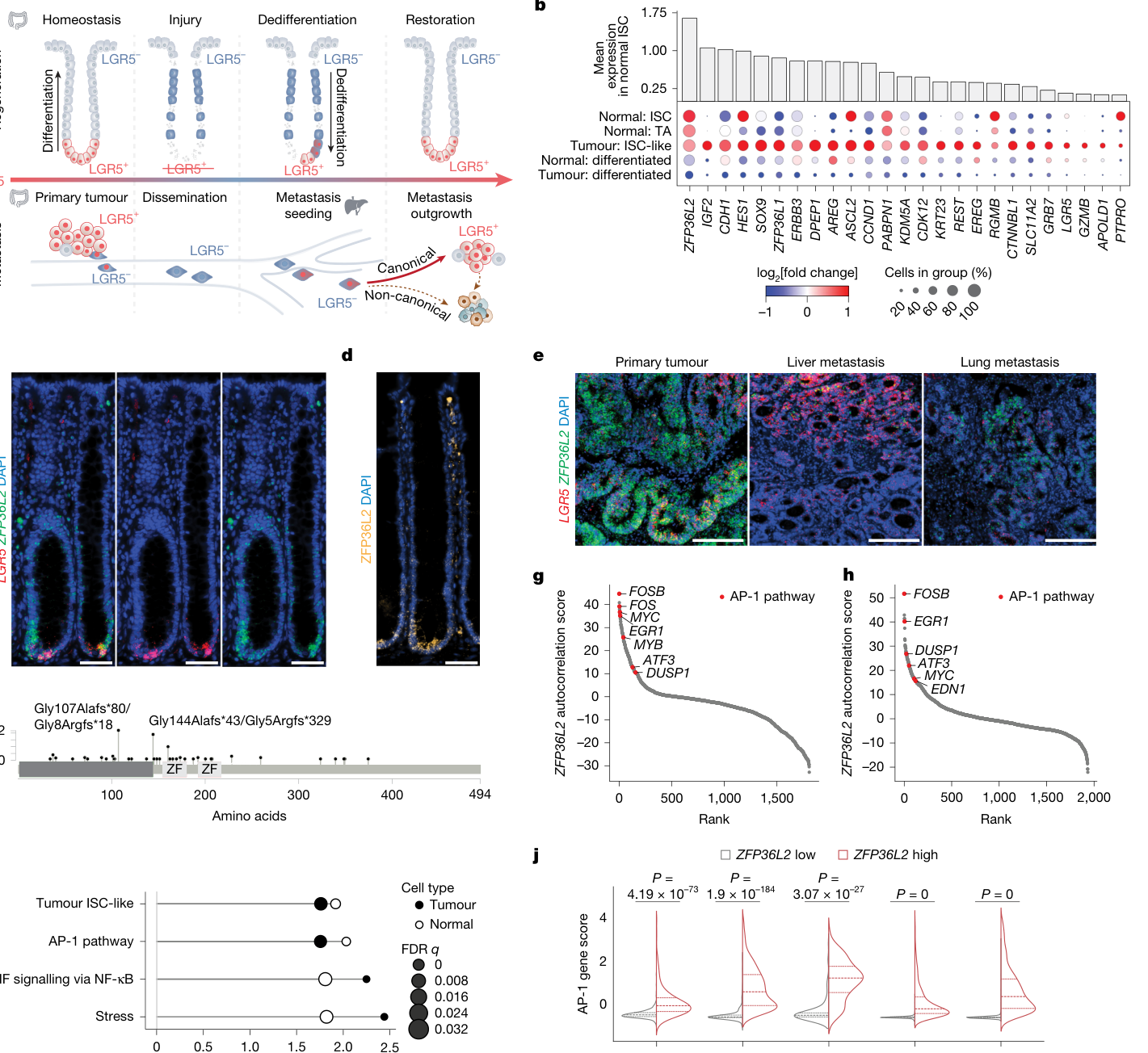
Fig. 1. ZFP36L2 is expressed by ISCs and is associated with the AP-1 gene program. a, Schematic of cell-state plasticity and dynamics during intestinal regeneration (top) and CRC (bottom). b, Dot plot of tumour ISC-like module10 gene expression in normal colon (Normal) and CRC (Tumour) epithelial cells from 25 patients. The dot size indicates the per cent cells (rows) expressing each gene (columns). Colour scale indicates the log2 fold change in average gene expression. Bar plots show the mean expression of each gene across normal colon ISCs. Genes meeting false discovery rate (FDR) < 0.05 using the Wilcoxon rank-sum test (ISC versus all other epithelial subtypes) are shown. c,d, Representative LGR5 and ZFP36L2 RNA FISH (c) and ZFP36L2 IF (d) images of human normal colon crypts. e, Representative LGR5 and ZFP36L2 RNA FISH images of human primary CRC tumours (n = 2), liver metastases (n = 2) and lung metastases (n = 2) from 2 donors. f, Lollipop plot showing ZFP36L2 mutation(图注取自PDF文本层,来源:Nature, 2026)
2. 体外敲掉它看表型:再生能力直接残了
接下来他们做最经典的"缺失实验"——肠上皮特异性敲除小鼠。ZFP36L2 敲除小鼠在稳态下几乎正常,隐窝形态、体重、生存都没大问题,只是 LGR5⁺ 干细胞数量略少。
但一旦用 DSS 诱导结肠炎(一种损伤-再生模型),差别就出来了:敲除小鼠体重恢复更慢、隐窝结构修复更差。再用 LGR5-DTR 系统直接干掉 LGR5⁺ 细胞,让剩余的 LGR5⁻ 细胞去再生——ZFP36L2 敲除后,再生出来的 LGR5⁺ 细胞显著减少。
@方法论点评:注意这里的实验设计:干性细胞直接缺失模型(LGR5-DTR)排除了"ZFP36L2 只是影响已有干细胞的存活"这个解释,直接证明它影响的是非干细胞向干细胞的重新编程。
为了进一步排除体内微环境的影响,他们拿敲除小鼠的结肠隐窝做成类器官——体外同样观察到去分化能力下降。
这确立了第一层结论:在正常组织里,ZFP36L2 不是日常运转必需,而是"损伤后重编程"的关键许可因子。
3. 那么问题来了:在癌症里,到底是"刹车"还是"油门"?
这就有意思了。他们在 CRC 类器官里敲低 ZFP36L2,然后分别打入小鼠盲肠(原位)和脾脏(模拟血行转移):
原位盲肠:用经典/肠道谱系的 CRC 类器官(MSK125P, OKG146P),敲低后肿瘤长得更慢;但用已经具有非经典分化潜能的转移来源类器官(OKG146Li),敲低对原发瘤生长没影响。 脾脏注射:不管哪种类器官,ZFP36L2 敲低都显著减少肝转移播种。BLI 信号在接种后前两周持续下降,说明转移细胞在最初的应激期大量死掉或无法启动生长。
@方法论点评:这里非常关键——表型依赖于"背景"。在原位(肠道微环境),ZFP36L2 缺失对"非经典"肿瘤影响不大;在转移(陌生、高应激微环境),它的缺失让癌细胞无法完成"应激-干性重编程"这条路径。这其实是"应激启动干性程序"这条通路在转移中被放大了。
那幸存下来的那些转移灶呢?它们 LGR5 表达下降,而 KRT20(经典分化)、CHGB(神经内分泌)、CK5(鳞状)表达升高。
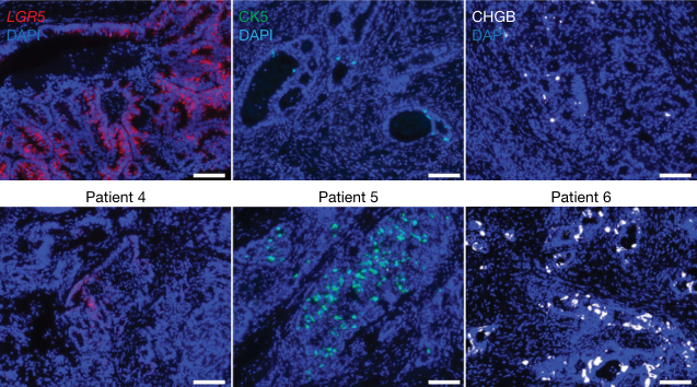
Fig. 3. Loss of ZFP36L2 inhibits CRC metastasis seeding and ISC dedifferentiation but promotes non-canonical differentiation. a–c, Orthotopic caecal xenotransplantation experiments. a, Schematic of the experiment. DOX, doxycycline. b, In vivo abdominal BLI average radiance, normalized to BLI at the time of doxycycline diet initiation. shL2, shZFP36L2. n (left to right) = 8, 5, 7, 8, 8, 14, 10 and 7 animals per group. Mean ± s.e.m.; two- tailed Mann–Whitney U-tests. c, End point ex vivo BLI of MSK107Li samples. Metastasis signals were normalized to orthotopic caecal signals in the same animals. n = 10 (shCtrl) and 7 (shL2) mice. Mean ± s.e.m.; two-tailed Mann– Whitney U-tests. d–g, Orthotopic liver metastasis seeding experiments. d, Schematic of the experiment. e, Representative ex vivo images of MSK107Li liver metastases. f, Average radiance of week 13 metastasis normalized to week 0 BLI. n = 6 (shCtrl) and 5 (shL2) mice. Mean ± s.e.m.; two-tailed Mann–Whitney U-tests. g, Weekly whole-body in vivo liver BLI (mean ± s.e.m.) normalized to week 0. n = 7 (shCtrl) and 5 (shL2) mice. Two-tailed Mann–Whitney U-tests. h, Schematic of the experiment. i. Kernel density estimate contour plots of scRNA-seq data from h, showing the overlap and divergence of cell states in OKG146P organoids transduced with shCtrl or shL2 and cultured in HISC, IGFF(图注取自PDF文本层,来源:Nature, 2026)
4. 机制层面:它怎么"感知"应激?——形成应激诱导的凝聚体
这里就是整篇最亮眼的部分。
他们发现内源性 ZFP36L2 在细胞里不是弥散的,而是呈斑点状分布。加上 AlphaFold 预测显示 ZFP36L2 有大量固有无序区(IDR)。这让人联想到"液-液相分离"凝聚体。
他们做了几个实验:
在活细胞里表达 ZFP36L2-eGFP,看到它形成动态、可移动的凝聚体。 临床常见的截短突变 fsZFP36L2 不能形成凝聚体。 FRAP 实验显示 ZFP36L2 凝聚体恢复缓慢(有相分离特征)。 加 RNase 或 1,6-己二醇(破坏相分离)都能让凝聚体消失,说明它依赖 RNA 和疏水作用。 撤除生长因子(应激)或化疗药能迅速诱导凝聚体形成,不依赖蛋白总量变化。
@方法论点评:这里用到了一个精巧的因果链条:“应激 → 凝聚体形成 → mRNA 降解 → 应激终止 → 干性重编程”。他们特意验证了"凝聚体数量峰值发生在蛋白总量峰值之前",证明应激诱导的是相分离行为本身,而不仅仅是蛋白表达上调。

Fig. 4. ZFP36L2 forms RNA-dependent, stress-responsive biomolecular condensates. a, Representative endogenous ZFP36L2 and pan-cytokeratin (Pan-CK) IF images of primary CRC and CRC liver metastasis from patient KG146. b, ZFP36L2 IF in MSK107Li organoids transduced with shCtrl (top) or shZFP36L2, demonstrating efficient knockdown and staining specificity. c,d, Top, AlphaFold2-predicted protein structure of human WT ZFP36L2 (c) and CRC patient hotspot mutation Gly144Alafs43 (d). Bottom, schematic of the protein structures indicating the CCCH zinc fingers (ZFs) and the N-terminal and C-terminal IDRs. Red, ZF domains (amino acids 153–219); grey, IDRs. e, Representative images of maximum-intensity projection of motion-artefact- corrected 3D time-lapse video of OKG146Li organoids co-expressing ZFP36L2– eGFP and H2B–mCherry. Top, condensate positions and trajectories overlaid on the original image. Bottom, corresponding trajectory rendering shown separately. f, Representative images of single z plane confocal live-cell microscopy of MSK107Li H2B–mCherry (red) organoids with doxycycline- inducible expression of eGFP, WT ZFP36L2–eGFP or fsZFP36L2–eGFP.(图注取自PDF文本层,来源:Nature, 2026)
5. 最后的拼图:凝聚体是拿来降解"应激 mRNA"的
他们用 HyperTRIBE(一种把 RNA 结合蛋白融合到 ADAR 酶的方法)在全转录组水平抓出 ZFP36L2 结合的 mRNA:几乎全是 3’UTR 含 AU-rich 元件的转录本,大量属于应激反应基因、缺氧、TNF、凋亡通路。
然后放两个实验交叉验证:
Actinomycin D 阻断新转录后,ZFP36L2 敲除后稳定性上升的 mRNA,正好重叠 HyperTRIBE 靶标。 SLAM-seq(4sU 代谢标记)直接测半衰期:ZFP36L2 敲除后,大量应激通路 mRNA 半衰期延长。
最后 FISH + IF 共定位看到:靶标 mRNA(如 GDF15、GADD45A)与 ZFP36L2 凝聚体共定位,急性应激时共定位下降(因为 mRNA 被快速降解了),而撤除应激后恢复。
@方法论点评:这一步最可贵的是"把凝聚体功能和 mRNA 降解动态联系在一起"——不是只证明"它们在一起",而是证明了"应激时凝聚体形成、mRNA 快速清除;应激解除后,凝聚体消退、mRNA 恢复"。这是动态因果的黄金标准。
核心结论
ZFP36L2 是应激-可塑性回路的核心开关:
应激感知:AP-1 应激通路激活,ZFP36L2 被诱导。 凝聚体形成:ZFP36L2 结合应激相关 mRNA,利用 IDR 和 RNA 支架效应形成动态凝聚体。 应激终止:凝聚体集中招募 mRNA 降解机器,迅速降解应激转录本,把 AP-1 驱动的"警报"消音。 干性重编程:应激终止后,细胞获得"许可"进入 LGR5⁺ 干性状态,再生或转移得以启动。
功能缺失时:应激无法被有效终止,细胞卡在非干性的应激状态;但在转移这种高选择性压力下,ZFP36L2⁻ 细胞被"迫"转向非经典分化(神经内分泌、鳞状)以求生存——这反而带来了更差的预后。
对耐药/DTP/PGCC 的启示
DTP 和 PGCC 的"应激刹车"可能依赖于类似机制。化疗本质上是强应激,药物耐受持久细胞(DTP)常表现出上皮-间质可塑性、应激颗粒上调。这篇提示:ZFP36L2 通路也许正是化疗后让 DTP"恢复干性"而再生的关键;缺失它,细胞可能直接走向非经典谱系(比如 PGCC 产生子细胞)。 “应激刹车"缺失可能驱动非经典耐药表型。临床上,化疗后耐药的 CRC 常出现神经内分泌分化。本文的"ZFP36L2 缺失 → 非经典分化"在机制上吻合。未来或许可以把 ZFP36L2 突变状态视为"是否容易发生谱系转换耐药"的生物标志物。 相分离凝聚体作为药物靶点的可能性。ZFP36L2 的凝聚体是依赖 RNA 的动态结构,传统小分子难以直接靶向 IDR。但如果能找到调控其凝聚体稳定性的上游激酶(如 MAPK/ERK 通路),可能干预"应激-干性"转换,而不影响正常稳态。
局限
本文的核心模型来自 CRC,虽然提到胰腺癌、黑色素瘤也有 ZFP36L2 突变,但未做跨癌种系统验证。 ZFP36L2 凝聚体的"降解应激 mRNA"是否必需其相分离能力,还是只是"伴随现象”,文中没有做"破坏相分离但不影响结合"的突变体实验来严格证明。 临床标本中 ZFP36L2 突变与预后的关联是基于小样本免疫组化和 FISH,未用大规模队列做多因素分析(文中主要是描述性统计)。 SLAM-seq 和 ActD 实验是在类器官里做的,没有在体内转移微环境里直接验证 mRNA 降解动力学。
来源
期刊:Nature,2026。DOI: 10.1038/s41586-026-10890-0。